. 2018 Aug;41(4):1157-1171.
doi: 10.1007/s10753-018-0763-1.

SERMs Promote Anti-Inflammatory Signaling and Phenotype of CD14+ Cells

Affiliations

SERMs Promote Anti-Inflammatory Signaling and Phenotype of CD14+ Cells

Lauri Polari et al. Inflammation. 2018 Aug.

Abstract

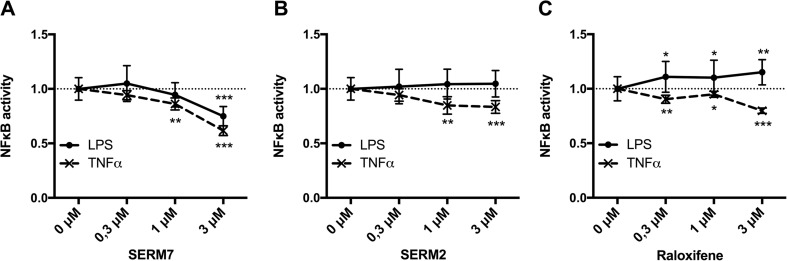
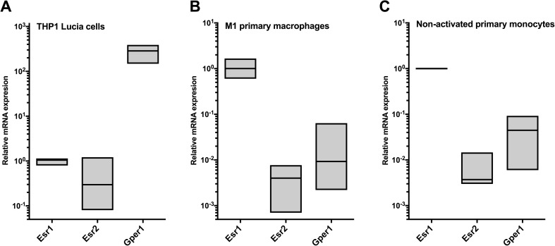
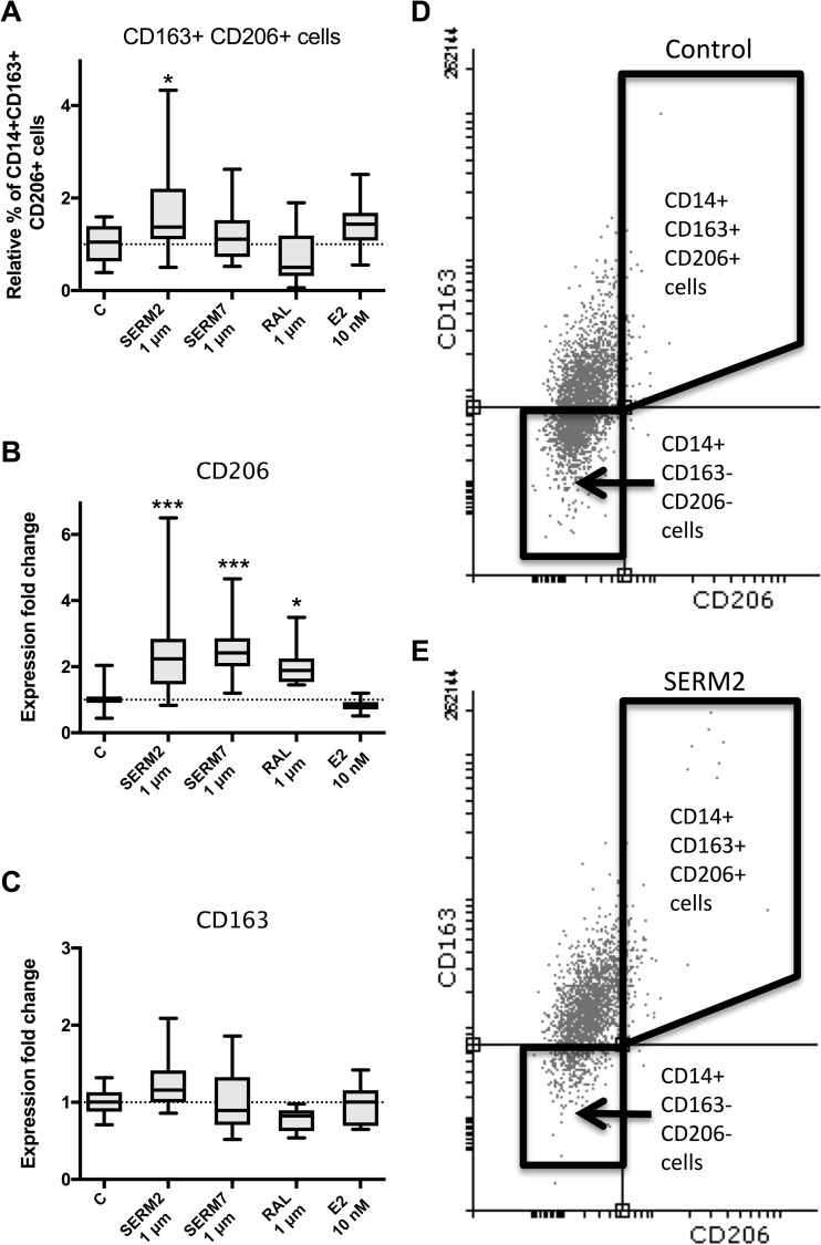
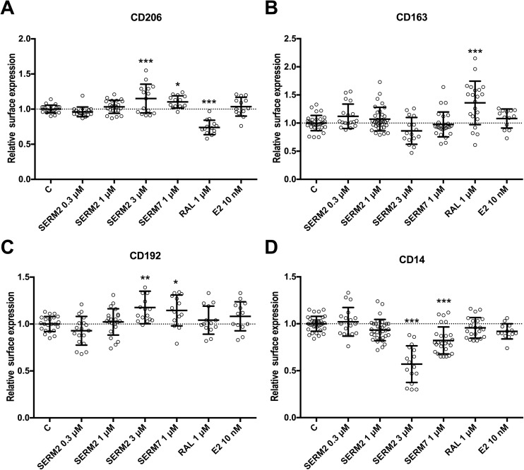
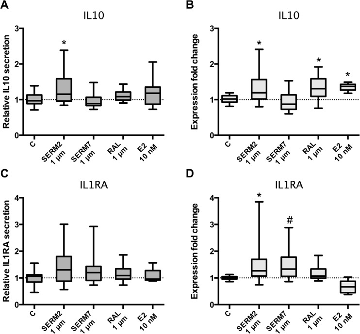
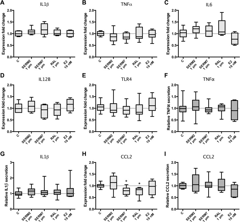
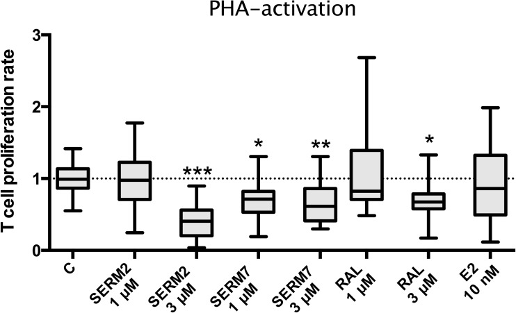
Signaling via estrogen receptors (ER) is recognized as an essential part of the immune regulation, and ER-mediated signaling is involved in autoimmune reactions. Especially ERα activation in immune cells has been suggested to skew cytokine production toward Th2/M2-type mediators, which can have protective effect on inflammatory diseases and reduce Th1 and Th17 responses. These effects are caused by increased alternative activation of macrophages and changes in the activation of different T cell populations. In humans, hormonal status has been shown to have a major impact on several inflammatory diseases. Selective estrogen receptor modulators (SERMs) are ER ligands that regulate ER actions in a tissue-specific manner mostly lacking the adverse effects of steroid hormones. The impact of SERMs on the immune system is less studied, but it is suggested that certain SERMs may also produce immunoprotective effects. Here, we show that two novel SERMs and raloxifene affect immune cells by promoting M2 macrophage phenotype, alleviating NFκB activity, inhibiting T cell proliferation, and stimulating the production of anti-inflammatory compounds such as IL10 and IL1 receptor antagonist. Thus, these compounds have high potency as drug candidates against autoimmune diseases.

Keywords: SERM; estradiol; estrogen receptor; inflammation; macrophages; raloxifene.


Conflict of interest statement

Lauri Kangas and Tero Linnanen were employees of Forendo Pharma Ltd. at the time of the studies and during the manuscript development.

Figures

Fig. 1
Fig. 1
SERMs modulate NFκB activity in monocytic THP-1 Lucia reporter cells. Effects of a SERM7, b SERM2, and c raloxifene on 24-h LPS or TNF-induced reporter activity of THP-1 monocytes. c(LPS) = 10 ng/ml, c(TNFα) = 5 ng/ml. Dots and error bars represent mean ±SD. Statistical significance was determined after unpaired t test vs control (0 μM). *P < 0.05; **P < 0.01; ***P < 0.001.
Fig. 2
Fig. 2
Expression of estrogen receptor subtypes (Esr1 and Esr2) and Gper1 genes in a THP-1 Lucia cells, b CD14-positive primary M(IFNγ+LPS) cells, and c non-activated monocytes. Boxes extend from min to max, and line in the middle represents median.
Fig. 3
Fig. 3
SERMs promote an alternative macrophage activation of monocytes by inducing activation of anti-inflammatory CD14+ CD163+ CD206 macrophage phenotype. a Proportion of CD14+ CD163+ CD206+ macrophages from CD14+ cell population. b Gene expression of CD206 and c CD163 in CD14+ M(IFNγ+LPS) cells. Box extends from the 25th to 75th percentiles; line in the middle represents median, and whisker from min to max. One-way statistical significance was determined after one-way ANOVA and Holm-Sidak post hoc multiple comparison test. *P < 0.05; **P < 0.01; ***P < 0.001. d, e Representative FACS dot plots (CD163-AF647 vs CD206-AF488) showing M1 (lower left quadrant) and double-positive CD163+ CD206+ (upper right quadrant) M2-like cells after IFNγ+LPS.
Fig. 4
Fig. 4
Effect of 48-h SERM/E2 treatment on median fluorescence intensity representing surface receptor expression of a CD206, b CD163, c CD192, and d CD14 in human-derived CD14-positive mononuclear cells, cultured 6 days for monocyte polarization and IFNγ 50 ng/ml followed by LPS activation at day 5. Scatter plot of individual samples with means ± SD. Statistical significance vs control group was determined after one-way ANOVA followed by post hoc Holm-Sidak multiple comparison test. *P < 0.05; **P < 0.01; ***P < 0.001.
Fig. 5
Fig. 5
Estrogen receptor ligands modulate cytokine synthesis and secretion of (IFNγ+LPS)-activated CD14+ cells toward anti-inflammatory phenotype. a Secretion and b gene expression rates of IL10. c, d Respective values for IL1RA. Five hundred thousand cells per sample. Box extends from the 25th to 75th percentiles and whiskers from min to max. Statistical significance was determined after one-way ANOVA followed by post hoc Holm-Sidak multiple comparison test. #P ≤ 0.1; *P < 0.05.
Fig. 6
Fig. 6
Estrogen receptor ligands do not downregulate proinflammatory signaling in (IFNγ+LPS)-activated CD14+ macrophages. Gene expression rates of a IL1β, b TNFα, c IL6, d IL12B, and e TLR4. Secretion rates of f TNFα and g IL1β from M(IFNγ+LPS) cells. h Gene expression and i secretion rates of CCL2. Five hundred thousand cells per sample. Box and whisker as min and max. Statistical significance was determined after one-way ANOVA followed by post hoc Holm-Sidak multiple comparison test. *P < 0.05.
Fig. 7
Fig. 7
SERMs inhibit proliferation of PHA-activated primary T cells. Peripheral blood-derived mononuclear cells from five donors were cultured 3 days with phytohemagglutinin (PHA) and SERM or E2. Proliferation rate of non-adherent cells was measured with β-counter after 20 h 3H-thymidine incorporation. Selective estrogen receptor modulator stimulates IL-10 secretion from IFNγ+LPS-activated CD14-positive monocytes. Box extends from the 25th to 75th percentiles; line in the middle represents median, and whisker from min to max. Statistical significance was determined after one-way ANOVA followed by post hoc Holm-Sidak multiple comparison test. *P < 0.05; **P < 0.01; ***P < 0.001.

References

    1. Vegeto E, Bonincontro C, Pollio G, et al. Estrogen prevents the lipopolysaccharide-induced inflammatory response in microglia. J Neurosci. 2001;21:1809–1818. doi: 10.1523/JNEUROSCI.21-06-01809.2001. - DOI - PMC - PubMed
    1. Polanczyk MJ, Carson BD, Subramanian S, et al. Cutting edge: estrogen drives expansion of the CD4+CD25+ regulatory T cell compartment. Journal of Immunology. 2004;173:2227–2230. doi: 10.4049/jimmunol.173.4.2227. - DOI - PubMed
    1. Saijo K, Collier JG, Li AC, et al. An ADIOL-ERb-CtBP transrepression pathway negatively regulates microglia-mediated inflammation. Cell. 2011;145:584–595. doi: 10.1016/j.cell.2011.03.050. - DOI - PMC - PubMed
    1. Lugo L, Villalvilla A, Largo R, et al. Selective estrogen receptor modulators (SERMs): new alternatives for osteoarthritis? Maturitas. 2014;77:380–384. doi: 10.1016/j.maturitas.2014.01.016. - DOI - PubMed
    1. Paterni I, Granchi C, Katzenellenbogen JA, Minutolo F. Estrogen receptors alpha (ERa) and beta (ERb): subtype-selective ligands and clinical potential. Steroids. 2014;90:13–29. doi: 10.1016/j.steroids.2014.06.012. - DOI - PMC - PubMed

Substances