

Cortical microstructure in young onset Alzheimer's disease using neurite orientation dispersion and density imaging
- PMID: 29575324
- PMCID: PMC6055830
- DOI: 10.1002/hbm.24056
Cortical microstructure in young onset Alzheimer's disease using neurite orientation dispersion and density imaging
Abstract
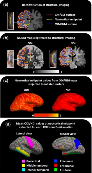
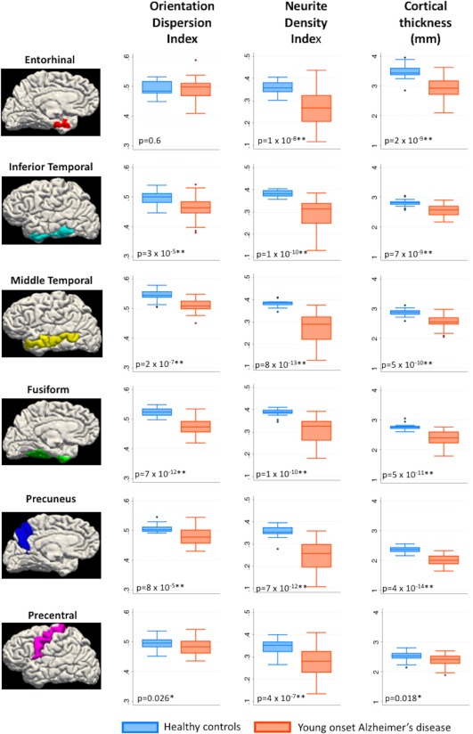
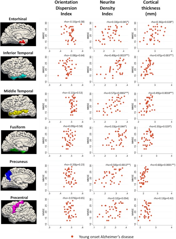
Alzheimer's disease (AD) is associated with extensive alterations in grey matter microstructure, but our ability to quantify this in vivo is limited. Neurite orientation dispersion and density imaging (NODDI) is a multi-shell diffusion MRI technique that estimates neuritic microstructure in the form of orientation dispersion and neurite density indices (ODI/NDI). Mean values for cortical thickness, ODI, and NDI were extracted from predefined regions of interest in the cortical grey matter of 38 patients with young onset AD and 22 healthy controls. Five cortical regions associated with early atrophy in AD (entorhinal cortex, inferior temporal gyrus, middle temporal gyrus, fusiform gyrus, and precuneus) and one region relatively spared from atrophy in AD (precentral gyrus) were investigated. ODI, NDI, and cortical thickness values were compared between controls and patients for each region, and their associations with MMSE score were assessed. NDI values of all regions were significantly lower in patients. Cortical thickness measurements were significantly lower in patients in regions associated with early atrophy in AD, but not in the precentral gyrus. Decreased ODI was evident in patients in the inferior and middle temporal gyri, fusiform gyrus, and precuneus. The majority of AD-related decreases in cortical ODI and NDI persisted following adjustment for cortical thickness, as well as each other. There was evidence in the patient group that cortical NDI was associated with MMSE performance. These data suggest distinct differences in cortical NDI and ODI occur in AD and these metrics provide pathologically relevant information beyond that of cortical thinning.
Keywords: Alzheimer's disease; dementia; diffusion MRI; grey matter; neurite orientation dispersion and density imaging; neurodegenerative disorders.
© 2018 The Authors Human Brain Mapping Published by Wiley Periodicals, Inc.
Figures
Similar articles
-
Association between diffusion MRI-based measures of neurite microstructure and risk of Alzheimer's disease.Exp Gerontol. 2025 Jul;206:112782. doi: 10.1016/j.exger.2025.112782. Epub 2025 May 14. Exp Gerontol. 2025. PMID: 40378932
-
Astrogliosis, neuritic microstructure, and sex effects: GFAP is an indicator of neuritic orientation in women.Brain Behav Immun. 2023 Oct;113:124-135. doi: 10.1016/j.bbi.2023.06.026. Epub 2023 Jun 30. Brain Behav Immun. 2023. PMID: 37394144 Free PMC article.
-
Cortical Microstructural Alterations in Mild Cognitive Impairment and Alzheimer's Disease Dementia.Cereb Cortex. 2020 May 14;30(5):2948-2960. doi: 10.1093/cercor/bhz286. Cereb Cortex. 2020. PMID: 31833550 Free PMC article.
-
Structural magnetic resonance imaging for the early diagnosis of dementia due to Alzheimer's disease in people with mild cognitive impairment.Cochrane Database Syst Rev. 2020 Mar 2;3(3):CD009628. doi: 10.1002/14651858.CD009628.pub2. Cochrane Database Syst Rev. 2020. PMID: 32119112 Free PMC article.
-
A systematic review of microstructural abnormalities in multiple sclerosis detected with NODDI and DTI models of diffusion-weighted magnetic resonance imaging.Magn Reson Imaging. 2023 Dec;104:61-71. doi: 10.1016/j.mri.2023.09.010. Epub 2023 Sep 27. Magn Reson Imaging. 2023. PMID: 37775062
Cited by
-
Interaction of amyloid and tau on cortical microstructure in cognitively unimpaired adults.Alzheimers Dement. 2022 Jan;18(1):65-76. doi: 10.1002/alz.12364. Epub 2021 May 13. Alzheimers Dement. 2022. PMID: 33984184 Free PMC article.
-
Neurite orientation dispersion and density imaging reveals white matter and hippocampal microstructure changes produced by Interleukin-6 in the TgCRND8 mouse model of amyloidosis.Neuroimage. 2019 Nov 15;202:116138. doi: 10.1016/j.neuroimage.2019.116138. Epub 2019 Aug 28. Neuroimage. 2019. PMID: 31472250 Free PMC article.
-
Neurite orientation dispersion and density imaging can detect presymptomatic axonal degeneration in the spinal cord of ALS mice.Funct Neurol. 2018 Jul/Sept;33(3):155-163. Funct Neurol. 2018. PMID: 30457969 Free PMC article.
-
Mediation Analysis with Multiple Exposures and Multiple Mediators.Stat Med. 2024 Nov 10;43(25):4887-4898. doi: 10.1002/sim.10215. Epub 2024 Sep 9. Stat Med. 2024. PMID: 39250913 Free PMC article.
-
Age-related differences in white matter microstructure measured by advanced diffusion MRI in healthy older adults at risk for Alzheimer's disease.Aging Brain. 2022 Jan 25;2:100030. doi: 10.1016/j.nbas.2022.100030. eCollection 2022. Aging Brain. 2022. PMID: 36908893 Free PMC article.
References
-
- Baloyannis, S. J. , Manolides, S. L. , & Manolides, L. S. (2011). Dendritic and spinal pathology in the acoustic cortex in Alzheimer's disease: Morphological estimation in Golgi technique and electron microscopy. Acta Oto‐Laryngologica, 131(6), 610–612. - PubMed
-
- Beck, J. , Pittman, A. , Adamson, G. , Campbell, T. , Kenny, J. , Houlden, H. , … Mead, S. (2014). Validation of next‐generation sequencing technologies in genetic diagnosis of dementia. Neurobiology of Aging, 35(1), 261–265. - PubMed
Publication types
MeSH terms
Grants and funding
LinkOut - more resources
Full Text Sources
Other Literature Sources
Medical
