

Spermidine promotes nucleus pulposus autophagy as a protective mechanism against apoptosis and ameliorates disc degeneration
- PMID: 29575654
- PMCID: PMC5980193
- DOI: 10.1111/jcmm.13586
Spermidine promotes nucleus pulposus autophagy as a protective mechanism against apoptosis and ameliorates disc degeneration
Abstract
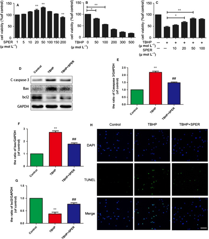
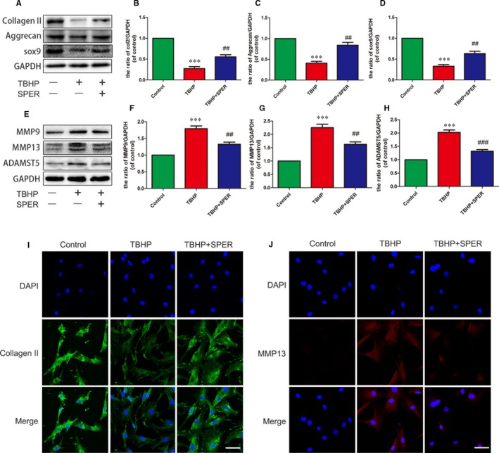
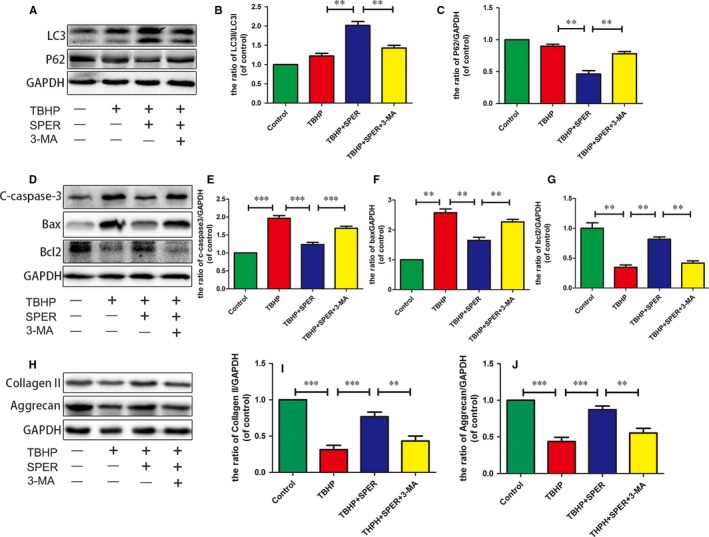
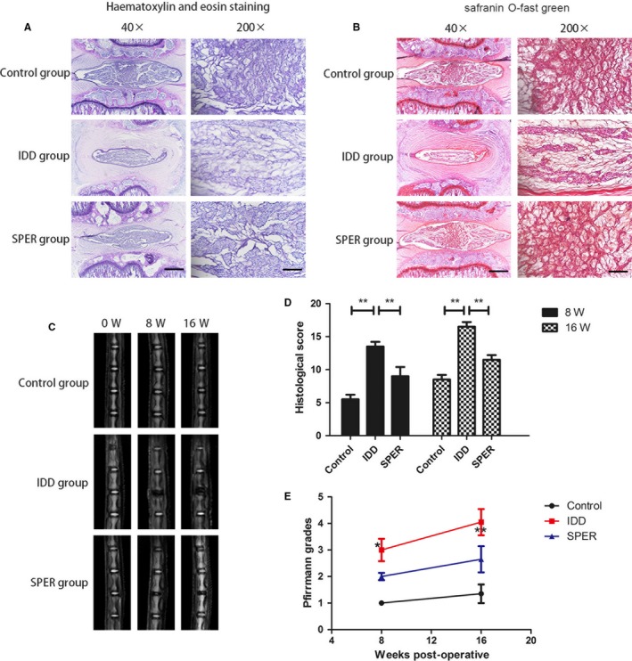
Spermidine has therapeutic effects in many diseases including as heart diastolic function, myopathic defects and neurodegenerative disorders via autophagy activation. Autophagy has been found to mitigate cell apoptosis in intervertebral disc degeneration (IDD). Accordingly, we theorize that spermidine may have beneficial effects on IDD via autophagy stimulation. In this study, spermidine's effect on IDD was evaluated in tert-butyl hydroperoxide (TBHP)-treated nucleus pulposus cells of SD rats in vitro as well as in a puncture-induced rat IDD model. We found that autophagy was actuated by spermidine in nucleus pulposus cells. In addition, spermidine treatment weakened the apoptotic effects of TBHP in nucleus pulposus cells. Spermidine increased the expression of anabolic proteins including Collagen-II and aggrecan and decreased the expression of catabolic proteins including MMP13 and Adamts-5. Additionally, autophagy blockade using 3-MA reversed the beneficial impact of spermidine against nucleus pulposus cell apoptosis. Autophagy was thus important for spermidine's therapeutic effect on IDD. Spermidine-treated rats had an accentuated T2-weighted signal and a diminished histological degenerative grade than vehicle-treated rats, showing that spermidine inhibited intervertebral disc degeneration in vivo. Thus, spermidine protects nucleus pulposus cells against apoptosis through autophagy activation and improves disc, which may be beneficial for the treatment of IDD.
Keywords: apoptosis; autophagy; intervertebral disc degeneration; oxidative stress; spermidine.
© 2018 The Authors. Journal of Cellular and Molecular Medicine published by John Wiley & Sons Ltd and Foundation for Cellular and Molecular Medicine.
Figures

Similar articles
-
Metformin protects against apoptosis and senescence in nucleus pulposus cells and ameliorates disc degeneration in vivo.Cell Death Dis. 2016 Oct 27;7(10):e2441. doi: 10.1038/cddis.2016.334. Cell Death Dis. 2016. PMID: 27787519 Free PMC article.
-
RTA 408 attenuates TBHP-Induced apoptosis in nucleus pulposus cells via Nrf2/ARE and NF-κB signaling pathways: in vitro and in vivo evidence for mitigating rats' intervertebral disc degeneration.Arthritis Res Ther. 2025 Jun 19;27(1):128. doi: 10.1186/s13075-025-03588-7. Arthritis Res Ther. 2025. PMID: 40537840 Free PMC article.
-
β‑ecdysterone protects against apoptosis by promoting autophagy in nucleus pulposus cells and ameliorates disc degeneration.Mol Med Rep. 2019 Mar;19(3):2440-2448. doi: 10.3892/mmr.2019.9861. Epub 2019 Jan 15. Mol Med Rep. 2019. PMID: 30664184
-
Circular RNAs in nucleus pulposus cell function and intervertebral disc degeneration.Cell Prolif. 2019 Nov;52(6):e12704. doi: 10.1111/cpr.12704. Epub 2019 Oct 16. Cell Prolif. 2019. PMID: 31621141 Free PMC article. Review.
-
MAPK /ERK signaling pathway: A potential target for the treatment of intervertebral disc degeneration.Biomed Pharmacother. 2021 Nov;143:112170. doi: 10.1016/j.biopha.2021.112170. Epub 2021 Sep 15. Biomed Pharmacother. 2021. PMID: 34536759 Review.
Cited by
-
Bushen Huoxue Formula Inhibits IL-1β-Induced Apoptosis and Extracellular Matrix Degradation in the Nucleus Pulposus Cells and Improves Intervertebral Disc Degeneration in Rats.J Inflamm Res. 2024 Jan 6;17:121-136. doi: 10.2147/JIR.S431609. eCollection 2024. J Inflamm Res. 2024. PMID: 38204990 Free PMC article.
-
Rebalance of the Polyamine Metabolism Suppresses Oxidative Stress and Delays Senescence in Nucleus Pulposus Cells.Oxid Med Cell Longev. 2022 Feb 7;2022:8033353. doi: 10.1155/2022/8033353. eCollection 2022. Oxid Med Cell Longev. 2022. PMID: 35178160 Free PMC article.
-
BMSC-Derived Exosomes Alleviate Intervertebral Disc Degeneration by Modulating AKT/mTOR-Mediated Autophagy of Nucleus Pulposus Cells.Stem Cells Int. 2022 Jul 9;2022:9896444. doi: 10.1155/2022/9896444. eCollection 2022. Stem Cells Int. 2022. PMID: 35855812 Free PMC article.
-
Autophagy Attenuates Oxidative Stress-Induced Collagen Degradation in Vaginal Fibroblasts: Implications for Pelvic Organ Prolapse.Int Urogynecol J. 2025 Mar;36(3):663-676. doi: 10.1007/s00192-024-06031-8. Epub 2025 Jan 23. Int Urogynecol J. 2025. PMID: 39847069
-
Polyphyllin I suppressed the apoptosis of intervertebral disc nucleus pulposus cells induced by IL-1β by miR-503-5p/Bcl-2 axis.J Orthop Surg Res. 2023 Jun 28;18(1):466. doi: 10.1186/s13018-023-03947-7. J Orthop Surg Res. 2023. PMID: 37380996 Free PMC article.
References
-
- Luoma K, Riihimaki H, Luukkonen R, et al. Low back pain in relation to lumbar disc degeneration. Spine. 2000;25:487‐492. - PubMed
-
- Stewart WF, Ricci JA, Chee E, et al. Lost productive time and cost due to common pain conditions in the US workforce. JAMA. 2003;290:2443‐2454. - PubMed
-
- Fontana G, See E, Pandit A. Current trends in biologics delivery to restore intervertebral disc anabolism. Adv Drug Deliv Rev. 2015;84:146‐158. - PubMed
-
- Raj PP. Intervertebral disc: anatomy‐physiology‐pathophysiology‐treatment. Pain Pract. 2008;8:18‐44. - PubMed
Publication types
MeSH terms
Substances
Grants and funding
LinkOut - more resources
Full Text Sources
Other Literature Sources
