

Clarification of the Innervation of the Bladder, External Urethral Sphincter and Clitoris: A Neuronal Tracing Study in Female Mongrel Hound Dogs
- PMID: 29575690
- PMCID: PMC6156994
- DOI: 10.1002/ar.23808
Clarification of the Innervation of the Bladder, External Urethral Sphincter and Clitoris: A Neuronal Tracing Study in Female Mongrel Hound Dogs
Abstract
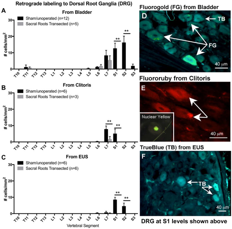
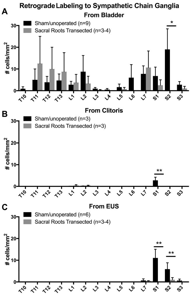
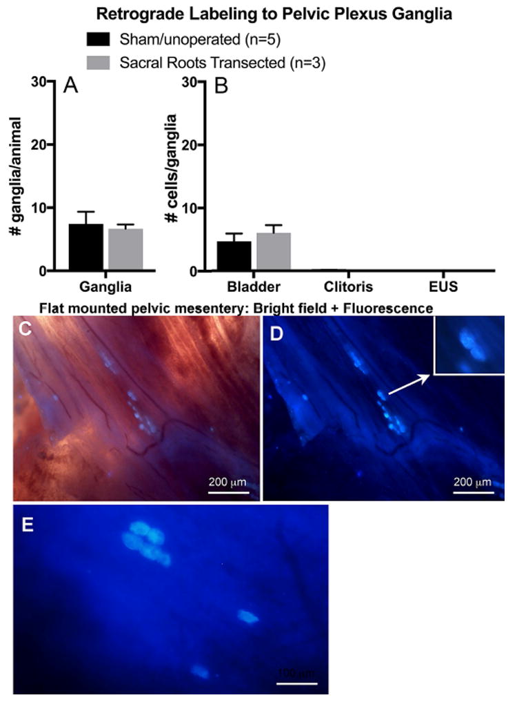
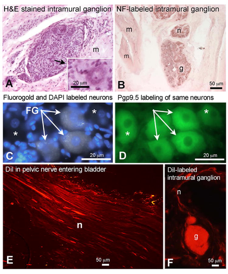
Many studies examining the innervation of genitourinary structures focus on either afferent or efferent inputs, or on only one structure of the system. We aimed to clarify innervation of the bladder, external urethral sphincter (EUS) and clitoris. Retrograde dyes were injected into each end organ in female dogs. Spinal cord, mid-bladder, and spinal, caudal mesenteric, sympathetic trunk and pelvic plexus ganglia were examined for retrograde dye-labeled neurons. Neurons retrogradely labeled from the bladder were found primarily in L7-S2 spinal ganglia, spinal cord lateral zona intermedia at S1-S3 levels, caudal mesenteric ganglia, T11-L2 and L6-S2 sympathetic trunk ganglia, and pelvic plexus ganglia. The mid-bladder wall contained many intramural ganglia neurons labeled anterogradely from the pelvic nerve, and intramural ganglia retrogradely labeled from dye labeling sites surrounding ureteral orifices. Neurons retrogradely labeled from the clitoris were found only in L7 and S1 spinal ganglia, L7-S3 spinal cord lateral zona intermedia, and S1 sympathetic trunk ganglia, and caudal mesenteric ganglia. Neurons retrogradely labeled from the EUS were found in primarily at S1 and S2 spinal ganglia, spinal cord lamina IX at S1-S3, caudal mesenteric ganglia, and S1-S2 sympathetic trunk ganglia. Thus, direct inputs from the spinal cord to each end organ were identified, as well as multisynaptic circuits involving several ganglia, including intramural ganglia in the bladder wall. Knowledge of this complex circuitry of afferent and efferent inputs to genitourinary structures is necessary to understand and treat genitourinary dysfunction. Anat Rec, 2018. © 2018 Wiley Periodicals, Inc.
Keywords: caudal mesenteric ganglia; clitoris; detrusor muscle; external urethral sphincter; spinal cord; spinal ganglion; sympathetic trunk ganglia.
© 2018 Wiley Periodicals, Inc.
Conflict of interest statement
Financial conflicts of interest, disclaimers: The authors have no financial conflicts of interest or disclaimers to declare.
Figures


Similar articles
-
Bladder reinnervation by somatic nerve transfer to pelvic nerve vesical branches does not reinnervate the urethra.Neurourol Urodyn. 2020 Jan;39(1):181-189. doi: 10.1002/nau.24217. Epub 2019 Nov 13. Neurourol Urodyn. 2020. PMID: 31724210 Free PMC article.
-
A quantitative analysis of the afferent and extrinsic efferent innervation of specific regions of the bladder and urethra in the cat.Brain Res Bull. 1984 Jun;12(6):735-40. doi: 10.1016/0361-9230(84)90154-0. Brain Res Bull. 1984. PMID: 6206931
-
Anatomical evidence for two spinal 'afferent-interneuron-efferent' reflex pathways involved in micturition in the rat: a 'pelvic nerve' reflex pathway and a 'sacrolumbar intersegmental' reflex pathway.Brain Res. 2000 Nov 10;883(1):107-18. doi: 10.1016/s0006-8993(00)02732-3. Brain Res. 2000. PMID: 11063993
-
Clitoral sexual arousal: neuronal tracing study from the clitoris through the spinal tracts.J Urol. 2008 Oct;180(4):1241-8. doi: 10.1016/j.juro.2008.06.009. Epub 2008 Aug 15. J Urol. 2008. PMID: 18707740 Free PMC article. Review.
-
Applied anatomy and physiology of the feline lower urinary tract.Vet Clin North Am Small Anim Pract. 1996 Mar;26(2):181-96. Vet Clin North Am Small Anim Pract. 1996. PMID: 8711856 Review.
Cited by
-
De novo aging-related NADPH diaphorase positive megaloneurites in the sacral spinal cord of aged dogs.Sci Rep. 2023 Dec 14;13(1):22193. doi: 10.1038/s41598-023-49594-0. Sci Rep. 2023. PMID: 38092874 Free PMC article.
-
A study on preganglionic connections and possible viscerofugal projections from urinary bladder intramural ganglia to the caudal mesenteric ganglion in the pig.J Anat. 2019 Feb;234(2):263-273. doi: 10.1111/joa.12916. Epub 2018 Nov 23. J Anat. 2019. PMID: 30468248 Free PMC article.
-
Mapping of spinal interneurons involved in regulation of the lower urinary tract in juvenile male rats.IBRO Rep. 2020 Jul 29;9:115-131. doi: 10.1016/j.ibror.2020.07.002. eCollection 2020 Dec. IBRO Rep. 2020. PMID: 32775758 Free PMC article.
-
Nerve transfer for restoration of lower motor neuron-lesioned bladder function. Part 1: attenuation of purinergic bladder smooth muscle contractions.Am J Physiol Regul Integr Comp Physiol. 2021 Jun 1;320(6):R885-R896. doi: 10.1152/ajpregu.00299.2020. Epub 2021 Mar 24. Am J Physiol Regul Integr Comp Physiol. 2021. PMID: 33759578 Free PMC article.
-
Aging-related NADPH diaphorase positive neurodegenerations in the sacral spinal cord of aged non-human primates.Sci Rep. 2024 Nov 8;14(1):27168. doi: 10.1038/s41598-024-77974-7. Sci Rep. 2024. PMID: 39511236 Free PMC article.
References
-
- Bahns E, Ernsberger U, Janig W, Nelke A. Functional characteristics of lumbar visceral afferent fibres from the urinary bladder and the urethra in the cat. Pflugers Arch. 1986;407:510–518. - PubMed
-
- Baljet B, Drukker J. The extrinsic innervation of the pelvic organs in the female rat. Acta Anat (Basel) 1980;107:241–267. - PubMed
-
- Barbe MF, Gomez-Amaya S, Braverman AS, Brown JM, Lamarre NS, Massicotte VS, Lewis JK, Dachert SR, Ruggieri MR., Sr Evidence of vagus nerve sprouting to innervate the urinary bladder and clitoris in a canine model of lower motoneuron lesioned bladder. Neurourol Urodyn. 2015;36(1):91–97. - PMC - PubMed
-
- Baron R, Janig W, McLachlan EM. The afferent and sympathetic components of the lumbar spinal outflow to the colon and pelvic organs in the cat. II. The lumbar splanchnic nerves. J Comp Neurol. 1985;238:147–157. - PubMed
Publication types
MeSH terms
Substances
Grants and funding
LinkOut - more resources
Full Text Sources
Other Literature Sources
