. 2018 May 14;57(20):5750-5753.
doi: 10.1002/anie.201801653. Epub 2018 Apr 14.

Fluorogenic Ag+ -Tetrazolate Aggregation Enables Efficient Fluorescent Biological Silver Staining

Affiliations

Fluorogenic Ag+ -Tetrazolate Aggregation Enables Efficient Fluorescent Biological Silver Staining

Sheng Xie et al. Angew Chem Int Ed Engl. .

Abstract

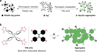
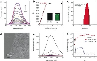
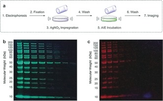
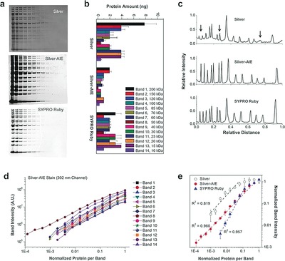
Silver staining, which exploits the special bioaffinity and the chromogenic reduction of silver ions, is an indispensable visualization method in biology. It is a most popular method for in-gel protein detection. However, it is limited by run-to-run variability, background staining, inability for protein quantification, and limited compatibility with mass spectroscopic (MS) analysis; limitations that are largely attributed to the tricky chromogenic visualization. Herein, we reported a novel water-soluble fluorogenic Ag+ probe, the sensing mechanism of which is based on an aggregation-induced emission (AIE) process driven by tetrazolate-Ag+ interactions. The fluorogenic sensing can substitute the chromogenic reaction, leading to a new fluorescence silver staining method. This new staining method offers sensitive detection of total proteins in polyacrylamide gels with a broad linear dynamic range and robust operations that rival the silver nitrate stain and the best fluorescent stains.

Keywords: aggregation-induced emission; metal-ion sensor; protein detection; silver staining; tetrazolate-silver assembly.


Conflict of interest statement

The authors declare no conflict of interest.

Figures

Figure 1
Figure 1
a) The classic chrome‐silver visualization (left) and proposed fluorogenic visualization (right) of proteins. b) TPE‐4TA and the Ag+ detection through a Ag+‐tetrazolate aggregation‐triggered AIE process. X can be either H or Na+. The possible tautomer 2X‐tetrazole structure (in red) is not shown for clarity.
Figure 2
Figure 2
Characterization of the fluorogenic aggregation process. a) Fluorescence of TPE‐4TA (5 μm) by stepwise addition of Ag+ in deionized water; b) Plot of intensity at 504 nm in (a) as a function of [Ag+]/[TPE‐4TA]. c) DLS and d) SEM characterization of the fluorescent solution. e) Fluorescence of TPE‐4TA (5 μm) mixed with metal ions (20 μm, including Na+, K+, Ca2+, Mg2+, Mn2+, Zn2+, Cu2+, Fe2+ Fe3+, Ni2+, Au+, Pb2+, Cd2+, Pd2+, and Co2+) in phosphate aqueous solution (pH 7.4). f) Fluorescence of TPE‐4TA (10 μm) at 504 nm against the pH values of the phosphate aqueous solution in the absence or presence of Ag+ or Hg2+ (10 equiv) respectively. λex=368 nm.
Figure 3
Figure 3
a) Procedure for fluorescent silver‐AIE staining . Gels stained by b) the silver‐AIE stain and c) a SYPRO Ruby stain, imaged in parallel under 365 nm irradiation.
Figure 4
Figure 4
Comparison of the silver nitrate, fluorescent silver‐AIE, and SYPRO Ruby staining methods. a) Representative gel images. In the lanes (from left to right), serial dilutions (2‐fold) of ladder proteins were loaded from 200–500 ng (1st lane) to 0.012–0.003 ng (15th lane). b) Lower limit of detection (n=3). c) Signal profiles of the 5th lane (10–25 ng/band) showing the differential protein detection. d) Signal intensity against the protein amount by the silver‐AIE stain (n=3). e) Signal intensity as a function of protein quantity for the band 8 protein (n=3), showing LDR of the three methods. X‐axis is the normalized amount of protein per band, where 1=200–500 ng/band.

References

    1. Grant G., Brain Res. Rev. 2007, 55, 490–498. - PubMed
    1. Uchihara T., Acta Neuropathol. 2007, 113, 483–499. - PMC - PubMed
    1. None
    1. Grocott R. G., Am. J. Clin. Pathol. 1955, 25, 975–979; - PubMed
    1. Procop G. W., Haddad S., Quinn J., Wilson M. L., Henshaw N. G., Reller L. B., Artymyshyn R. L., Katanik M. T., Weinstein M. P., J. Clin. Microbiol. 2004, 42, 3333–3335. - PMC - PubMed

Publication types