

(Pro)renin receptor: Involvement in diabetic retinopathy and development of molecular targeted therapy
- PMID: 29575757
- PMCID: PMC6319493
- DOI: 10.1111/jdi.12842
(Pro)renin receptor: Involvement in diabetic retinopathy and development of molecular targeted therapy
Abstract
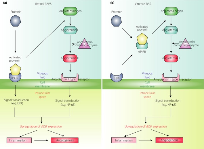
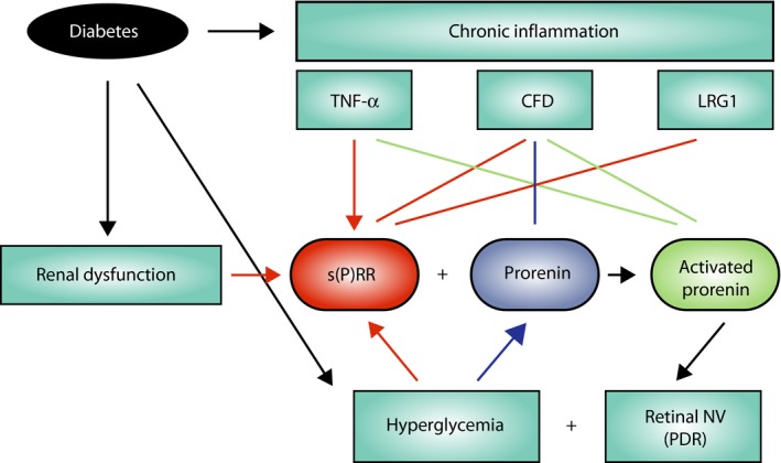
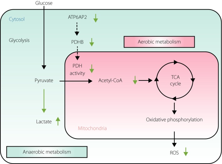
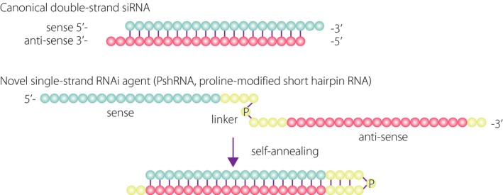
The renin-angiotensin system (RAS), a crucial regulator of systemic blood pressure (circulatory RAS), plays distinct roles in pathological angiogenesis and inflammation in various organs (tissue RAS), such as diabetic microvascular complications. Using ocular clinical samples and animal disease models, we elucidated molecular mechanisms in which tissue RAS excites the expression of vascular endothelial growth factor (VEGF)-A responsible for retinal inflammation and angiogenesis, the two major pathological events in diabetic retinopathy (DR). Furthermore, we showed the involvement of (pro)renin receptor [(P)RR] in retinal RAS activation and its concurrent intracellular signal transduction (e.g., extracellular signal-regulated kinase); namely, the (P)RR-induced dual pathogenic bioactivity referred to as the receptor-associated prorenin system. Indeed, neovascular endothelial cells in the fibrovascular tissue collected from eyes with proliferative DR were immunoreactive for the receptor-associated prorenin system components including prorenin, (P)RR, phosphorylated extracellular signal-regulated kinase and VEGF-A. Protein levels of soluble (P)RR increased with its positive correlations with prorenin, renin enzymatic activity and VEGF in the vitreous of proliferative DR eyes, suggesting a close link between (P)RR and VEGF-A-driven angiogenic activity. Furthermore, we revealed an unsuspected, PAPS-independent role of (P)RR in glucose-induced oxidative stress. Recently, we developed an innovative single-strand ribonucleic acid interference molecule selectively targeting human and mouse (P)RR, and confirmed its efficacy in suppressing diabetes-induced retinal inflammation in mice. Our data using clinical samples and animal models suggested the significant implication of (P)RR in the pathogenesis of DR, and the potential usefulness of the ribonucleic acid interference molecule as a therapeutic agent to attenuate ocular inflammation and angiogenesis.
Keywords: (Pro)renin receptor; Diabetic retinopathy; Receptor-associated prorenin system.
© 2018 The Authors. Journal of Diabetes Investigation published by Asian Association for the Study of Diabetes (AASD) and John Wiley & Sons Australia, Ltd.
Figures
Similar articles
-
(Pro)renin receptor is associated with angiogenic activity in proliferative diabetic retinopathy.Diabetologia. 2012 Nov;55(11):3104-13. doi: 10.1007/s00125-012-2702-2. Epub 2012 Aug 30. Diabetologia. 2012. PMID: 22930161
-
[(Pro) renin receptor in the pathogenesis of proliferative diabetic retinopathy].Nippon Ganka Gakkai Zasshi. 2014 Nov;118(11):916-26. Nippon Ganka Gakkai Zasshi. 2014. PMID: 25543383 Review. Japanese.
-
Receptor-associated prorenin system contributes to development of inflammation and angiogenesis in proliferative diabetic retinopathy.Inflamm Regen. 2016 Sep 7;36:22. doi: 10.1186/s41232-016-0027-0. eCollection 2016. Inflamm Regen. 2016. PMID: 29259695 Free PMC article. Review.
-
Combined renin inhibition/(pro)renin receptor blockade in diabetic retinopathy--a study in transgenic (mREN2)27 rats.PLoS One. 2014 Jun 26;9(6):e100954. doi: 10.1371/journal.pone.0100954. eCollection 2014. PLoS One. 2014. PMID: 24968134 Free PMC article.
-
(Pro)renin receptor-mediated signal transduction and tissue renin-angiotensin system contribute to diabetes-induced retinal inflammation.Diabetes. 2009 Jul;58(7):1625-33. doi: 10.2337/db08-0254. Epub 2009 Apr 23. Diabetes. 2009. PMID: 19389828 Free PMC article.
Cited by
-
Effects of Angiotensin Receptor Blockers on Streptozotocin-Induced Diabetic Cataracts.J Clin Med. 2023 Oct 19;12(20):6627. doi: 10.3390/jcm12206627. J Clin Med. 2023. PMID: 37892765 Free PMC article.
-
Receptor-Associated Prorenin System in the Trabecular Meshwork of Patients with Primary Open-Angle Glaucoma and Neovascular Glaucoma.J Clin Med. 2020 Jul 22;9(8):2336. doi: 10.3390/jcm9082336. J Clin Med. 2020. PMID: 32707887 Free PMC article.
-
Targeting aging and age-related diseases with vaccines.Nat Aging. 2024 Apr;4(4):464-482. doi: 10.1038/s43587-024-00597-0. Epub 2024 Apr 15. Nat Aging. 2024. PMID: 38622408 Review.
-
Local ocular renin-angiotensin-aldosterone system: any connection with intraocular pressure? A comprehensive review.Ann Med. 2020 Aug;52(5):191-206. doi: 10.1080/07853890.2020.1758341. Epub 2020 Apr 30. Ann Med. 2020. PMID: 32308046 Free PMC article.
-
Diabetic Retinopathy: A Pharmacological Consideration.Cureus. 2023 Oct 11;15(10):e46842. doi: 10.7759/cureus.46842. eCollection 2023 Oct. Cureus. 2023. PMID: 37954772 Free PMC article. Review.
References
-
- Ishida S, Usui T, Yamashiro K, et al VEGF164 is proinflammatory in the diabetic retina. Invest Ophthalmol Vis Sci 2003; 44: 2155–2162. - PubMed
-
- Adamis AP, Miller JW, Bernal MT, et al Increased vascular endothelial growth factor levels in the vitreous of eyes with proliferative diabetic retinopathy. Am J Ophthalmol 1994; 118: 445–450. - PubMed
-
- Aiello LP, Avery RL, Arrigg PG, et al Vascular endothelial growth factor in ocular fluid of patients with diabetic retinopathy and other retinal disorders. N Engl J Med 1994; 331: 1480–1487. - PubMed
-
- Malecaze F, Clamens S, Simorre‐Pinatel V, et al Detection of vascular endothelial growth factor messenger RNA and vascular endothelial growth factor‐like activity in proliferative diabetic retinopathy. Arch Ophthalmol 1994; 112: 1476–1482. - PubMed
-
- Hurwitz H, Fehrenbacher L, Novotny W, et al Bevacizumab plus irinotecan, fluorouracil, and leucovorin for metastatic colorectal cancer. N Engl J Med 2004; 350: 2335–2342. - PubMed
Publication types
MeSH terms
Substances
Grants and funding
LinkOut - more resources
Full Text Sources
Other Literature Sources
Medical
