. 2018 Apr 5;173(2):443-455.e12.
doi: 10.1016/j.cell.2018.02.047. Epub 2018 Mar 22.

Common PIEZO1 Allele in African Populations Causes RBC Dehydration and Attenuates Plasmodium Infection

Affiliations

Common PIEZO1 Allele in African Populations Causes RBC Dehydration and Attenuates Plasmodium Infection

Shang Ma et al. Cell. .

Abstract

Hereditary xerocytosis is thought to be a rare genetic condition characterized by red blood cell (RBC) dehydration with mild hemolysis. RBC dehydration is linked to reduced Plasmodium infection in vitro; however, the role of RBC dehydration in protection against malaria in vivo is unknown. Most cases of hereditary xerocytosis are associated with gain-of-function mutations in PIEZO1, a mechanically activated ion channel. We engineered a mouse model of hereditary xerocytosis and show that Plasmodium infection fails to cause experimental cerebral malaria in these mice due to the action of Piezo1 in RBCs and in T cells. Remarkably, we identified a novel human gain-of-function PIEZO1 allele, E756del, present in a third of the African population. RBCs from individuals carrying this allele are dehydrated and display reduced Plasmodium infection in vitro. The existence of a gain-of-function PIEZO1 at such high frequencies is surprising and suggests an association with malaria resistance.

Keywords: PIEZO1; cerebral malaria; dehydration; functional variants; genomics; human genetics; ion channel; malaria; mechanotransduction; red blood cell.


Conflict of interest statement

Declaration of Interests.

The authors declare no competing financial interests.

Figures

Figure 1
Figure 1
Mouse model for human xerocytosis. (A) Representative traces of mechanically-activated (MA) inward currents for wild type and Piezo1 R2482H. ****p < 0.001. (B) Strategy for generating knock-in mouse. (C) Osmotic fragility test for RBCs. (D) Quantification for osmotic fragility. Relative tonicity at which 50% RBCs are lysed (half hemolysis) was calculated for each curve. ****p < 0.001. (E) Scanning electron microcopy images. Heterozygous Piezo1GOF blood RBCs showed signs of stomatocytes. (F) Splenomegaly in gain-of-function Piezo1 mice (Fig S1C). Scale bar: 10μm. Data are presented as means ± SEM. See also Figure S1 and Table S1
Figure 2
Figure 2
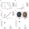
Plasmodium infection in gain-of-function Piezo1 mice. (A) Survival curves for gain-of-function Piezo1 mice after P. berghei infected RBCs (B) and (C) Parasitemia recorded by flow cytometry for phase 1 (first 7 days) and phase 1 and 2 together (24) days respectively. (D) Intact blood-brain barrier in infected gain-of-function Piezo1 mice. (E) Quantification of blood-brain barrier disruption. (F) Brain water content in infected brains. (*p<0.05, **p<0.01, and ***p<0.001). Scale bar: 5mm. Data are presented as means ± SEM.
Figure 3
Figure 3
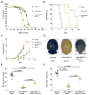
Role of RBC dehydration in Plasmodium infection in mice. (A) Deletion of KCa3.1 in heterozygous Piezo1GOF blood mice (orange) restored RBC dehydration in heterozygous Piezo1GOF blood (green). Piezo1GOF blood/ KCa3.1−/− mice had a similar survival curve to wild type. (B) Post infection survival rate of Piezo1GOF blood/ KCa3.1−/− mice (orange) is intermediate between wild type (black) and heterozygous Piezo1GOF blood mice (green). p<0.0001, (Mantel-Cox tests). (C) Both Piezo1GOF blood/ KCa3.1−/− and KCa3.1−/− mice had same parasitemia as wild type, with significantly higher than heterozygous Piezo1GOF blood mice. (D) Breakdown of blood-brain barrier in Piezo1GOF blood/ KCa3.1−/− mice 13 days after infection. (E) Quantification for blood-brain barrier disruption. (F) Brain water content in infected brains. (*p<0.05, **p<0.01, and ***p<0.001). Scale bar: 5mm. Data are presented as means ± SEM. See also Figure S2.
Figure 4
Figure 4
Role of gain-of-function Piezo1 expression in RBCs and T cells during Plasmodium infection in mice. (A) RBC osmotic fragility for different gain-of-function Piezo1 mice. (B) Mice with gain-of-function Piezo1 in different blood cells had distinct survival rates after infection. Piezo1GOF RBC (red) had survival rate similar to pan-blood cell-specific mice (Piezo1GOF blood (green)), p>0.05. Macrophage-specific gain-of-function mice (Piezo1GOF macrophage) had same survival rate as wild type, p>0.05. Piezo1GOF T-cells had a survival rate greater than wild type (p<0.01) and less than Piezo1GOF blood mice (p<0.01). Mantel-Cox tests. (C) Parasitemia recorded by flow-cytometry for gain-of-function mice. *p<0.05, **p<0.01, and ***p<0.001, student t-test. (D) Blood-brain barrier compromise in T cell- and RBC-specific gain-of-function mice after infection. (E) Quantification for blood-brain barrier disruption. (F) Brain water content in infected brains. Scale bar: 5mm. Data are presented as means ± SEM. See also Figure S3.
Figure 5
Figure 5
Identification of gain-of-function PIEZO1 mutations in African populations. (A) and (B) Yoda1 induced Intracellular calcium signals in PIEZO1KO HEK cells overexpressing A1988V and E756del cDNA (* p<0.05). Allele frequency for both mutations is shown in the insets. (C) Representative traces of mechanically activated (MA) inward currents for wild type and mutated cDNA. R2456H, A1988V, and E756del mutations. (D) Quantification for inactivation time (τ). n = number of cells. (***p < 0.001, **p < 0.01). Data are presented as means ± SEM
Figure 6
Figure 6
Population genetics of PIEZO1 gain-of-function E756del allele common in populations of African descent. (A) Human population demographics for E756 indels. E756 deletion (TCC/-) exists at high frequencies in all populations of African descents (purple). A minor allele, E756 insertion (TCC/TCCTCC) was also discovered (coral). (B) Differentiation (FST) between populations of African and non-African ancestry at each loci for all PIEZO1 missense mutations. Alleles are colored by whether the minor allele frequency (MAF) was highest in African (red) or non-African (black) populations, or were similar (grey). (C) A nucleotide alignment of modern and pre-modern (Neandertal and Denisovan) human PIEZO1 minus strand sequences around the E756del allele showing the codon positions. The TCC deletion (GGA on minus strand) spans two codons, but only deletes E756 while shifting nucleotides to leave D757 intact. Individual Neandertal and Denisovan reads used to create this alignment and comparisons to non-human primate PIEZO1 amino acid sequences are shown in Figure S4.
Figure 7
Figure 7
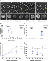
Characterization of RBCs from E756del carriers for xerocytosis-like phenotypes, and P. falciparum infection. (A) SEM images. Three individual E756del carriers have RBCs with echinocytes (white arrowhead) and stomatocytes (yellow arrowhead), magnified in lower panels. Scale bar for upper panels, 10um; for lower panels, 5um. (B) and (C) Osmotic fragility test. RBCs from E756del heterozygous carriers (n=9) had a left-shifted curve (blue) compared to controls (n=16) (black), as quantified in (C) **p <0.01. (D) and (E) P. falciparum infection into RBCs from E756del carriers. Giemsa staining (D) and SYBR Green labeling of parasite DNA inside RBCs (E) (**p < 0.01, *p < 0.05). Statistics: student’s t-test for each time point. Scale bar: 8mm for upper panels in A; 5mm for lower panels in A. Data are presented as means ± SEM. See also Figure S5.

References

    1. Albuisson J, Murthy SE, Bandell M, Coste B, Louis-Dit-Picard H, Mathur J, Fénéant-Thibault M, Tertian G, de Jaureguiberry JP, Syfuss PY, et al. Dehydrated hereditary stomatocytosis linked to gain-of-function mutations in mechanically activated PIEZO1 ion channels. Nat Commun. 2013;4:1884. - PMC - PubMed
    1. Archer NM, Shmukler BE, Andolfo I, Vandorpe DH, Gnanasambandam R, Higgins JM, Rivera A, Fleming MD, Sachs F, Gottlieb PA, et al. Hereditary xerocytosis revisited: Hereditary Xerocytosis Revisited. American Journal of Hematology. 2014;89:1142–1146. - PMC - PubMed
    1. Auton A, Brooks LD, Durbin RM, Garrison EP, Kang HM, Korbel JO, Marchini JL, McCarthy S, McVean GA, Abecasis GR with 1000 Genomes Project Consortium. A global reference for human genetic variation. Nature. 2015;526:68–74. - PMC - PubMed
    1. Bae C, Gnanasambandam R, Nicolai C, Sachs F, Gottlieb PA. Xerocytosis is caused by mutations that alter the kinetics of the mechanosensitive channel PIEZO1. Proc Natl Acad Sci USA. 2013;110:E1162–E1168. - PMC - PubMed
    1. Baptista FG, Pamplona A, Pena AC, Mota MM, Pied S, Vigário AM. Accumulation of Plasmodium berghei-infected red blood cells in the brain is crucial for the development of cerebral malaria in mice. Infect Immun. 2010a;78:4033–4039. - PMC - PubMed

Publication types

MeSH terms

Substances

Supplementary concepts