. 2018 Apr 2;28(7):1124-1131.e3.
doi: 10.1016/j.cub.2018.02.039. Epub 2018 Mar 22.

Mammalian Oocytes Locally Remodel Follicular Architecture to Provide the Foundation for Germline-Soma Communication

Affiliations

Mammalian Oocytes Locally Remodel Follicular Architecture to Provide the Foundation for Germline-Soma Communication

Stephany El-Hayek et al. Curr Biol. .

Abstract

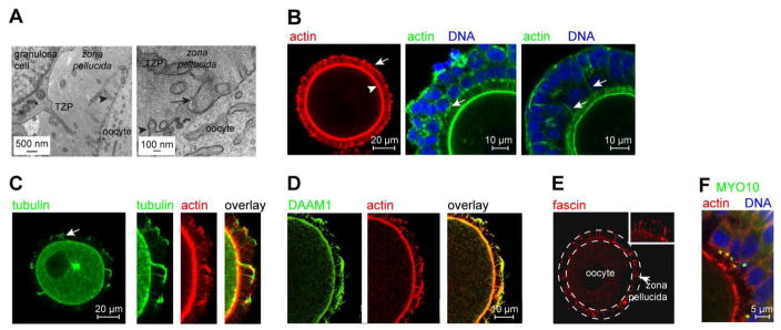
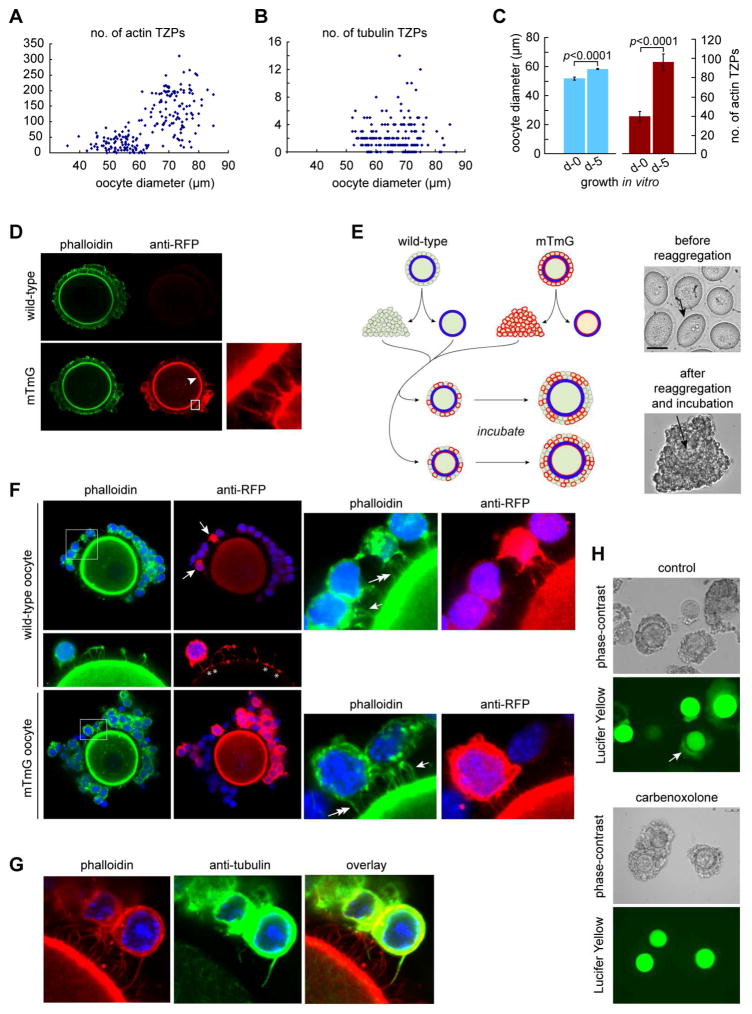
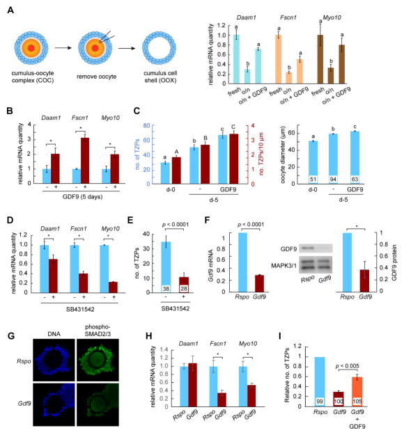
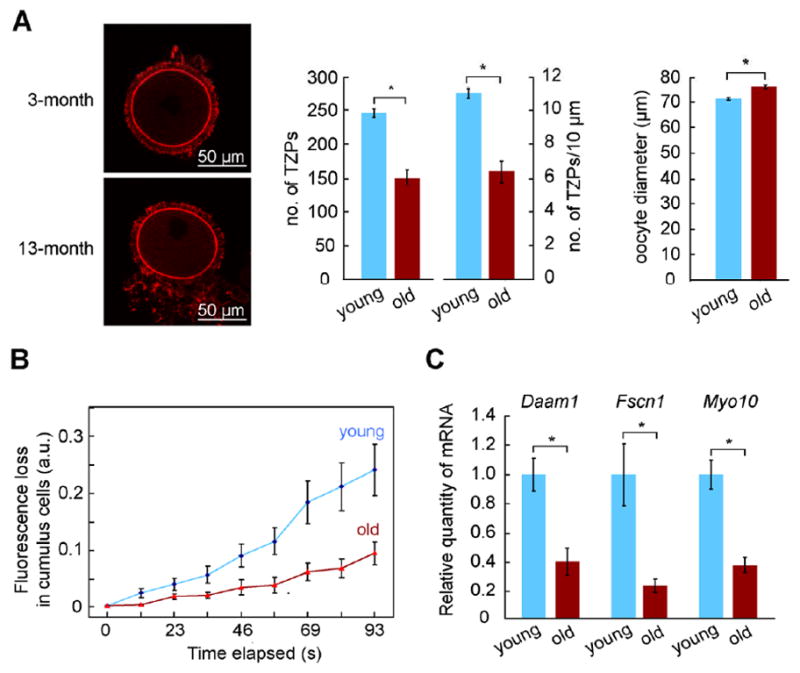
Germ cells develop in a microenvironment created by the somatic cells of the gonad [1-3]. Although in males, the germ and somatic support cells lie in direct contact, in females, a thick extracellular coat surrounds the oocyte, physically separating it from the somatic follicle cells [4]. To bypass this barrier to communication, narrow cytoplasmic extensions of the follicle cells traverse the extracellular coat to reach the oocyte plasma membrane [5-9]. These delicate structures provide the sole platform for the contact-mediated communication between the oocyte and its follicular environment that is indispensable for production of a fertilizable egg [8, 10-15]. Identifying the mechanisms underlying their formation should uncover conserved regulators of fertility. We show here in mice that these structures, termed transzonal projections (TZPs), are specialized filopodia whose number amplifies enormously as oocytes grow, enabling increased germ-soma communication. By creating chimeric complexes of genetically tagged oocytes and follicle cells, we demonstrate that follicle cells elaborate new TZPs that push through the extracellular coat to reach the oocyte surface. We further show that growth-differentiation factor 9, produced by the oocyte, drives the formation of new TZPs, uncovering a key yet unanticipated role for the germ cell in building these essential bridges of communication. Moreover, TZP number and germline-soma communication are strikingly reduced in reproductively aged females. Thus, the growing oocyte locally remodels follicular architecture to ensure that its developmental needs are met, and an inability of somatic follicle cells to respond appropriately to oocyte-derived cues may contribute to human infertility.

Keywords: fertility; filopodia; follicle; intercellular signaling; oocyte.


Conflict of interest statement

DECLARATION OF INTERESTS

The authors declare no competing interests

Figures

Figure 1
Figure 1. TZPs are specialized filopodia
(A) Left: TZP emanating from a granulosa cell. Arrowhead indicates an oocyte microvillus. Right: TZP reaching the oocyte. Arrow indicates bulbous ‘foot’. (B) Left: Oocyte stained using the actin-binding dye, phalloidin. The granulosa cell bodies have been removed to improve the resolution of the TZPs (arrow), which remain embedded in the zona pellucida. Arrowhead indicates the oocyte cortex. Middle, Right: GOCs stained using phalloidin. Middle: Multiple TZPs extend from each granulosa cell, often apparently from a single origin (arrow). Right: Actin-rich filaments (arrows) sometimes extend from peripheral layers of granulosa cells to the oocyte. (C) Oocyte stained using anti-tubulin and phalloidin. Left: Arrow indicates tubulin-rich TZP. Right: Some tubulin-TZPs also contain actin. (D) DAAM1 is present in TZPs. (E) Fascin is present in TZPs. The zona pellucida is traced by the dashed lines. Inset shows higher magnification. (F) MYO10 foci are present on the apical side of granulosa cells adjacent to the zona pellucida. No foci are detected in peripheral layers.
Figure 2
Figure 2. Granulosa cells elaborate new TZPs during oocyte growth
(A) Oocytes at different stages of growth were stained using phalloidin and the number of TZPs in an equatorial confocal optical section was counted. (B) As in (A) except that the oocytes were stained using anti-tubulin. (C) Oocyte diameter and number of actin-TZPs were determined in GOCs immediately after isolation or after 5 days of growth in vitro. (D) GOCs of wild-type (upper) or mTmG (lower) mice were stained using phalloidin and anti-RFP. Anti-RFP stains TZPs (inset) as well as the oocyte membrane (arrowhead) of mTmG mice. (E) Left: Reaggregation strategy. Right, upper: oocytes prior to reaggregation. Arrow indicates zona pellucida. Right, lower: reaggregated complex after 5 days of incubation. Arrow indicates oocyte. (F) Reaggregated complexes after incubation. Some granulosa cells have been removed to enable TZPs to be seen more clearly. Upper: Wild-type oocyte, whose membrane is not stained by anti-RFP, enclosed by wild-type and mTmG (arrows) granulosa cells. Right panel shows enlargement of the boxed area. TZPs of wild-type granulosa cells are stained by phalloidin only (single arrow); TZPs of mTmG granulosa cells are also stained by anti-RFP (double arrow). Middle: Asterisks illustrate where TZPs of mTmG granulosa cells contact the surface of a wild-type oocyte. Lower: mTmG oocyte, whose membrane is stained by anti-RFP, enclosed by wild-type and mTmG granulosa cells. Right panel shows enlargement of the boxed area. Single arrow and double arrows indicate TZPs as above. (G) TZP stained by both phalloidin and anti-tubulin. (H) Lucifer Yellow was injected into the oocyte of reaggregated complexes. Upper: bright-field; lower: dark-field. Arrow shows fluorescence in granulosa cells. Lower paired panels show complexes incubated in the gap junction blocker, carbenoxolone. See also Figure S1.
Figure 3
Figure 3. Oocyte-derived GDF9 promotes generation of new TZPs
(A) Left: Oocytectomy procedure. Oocytes were removed from COCs and the cumulus cell shells were harvested immediately (fresh) or cultured overnight on the absence or presence of GDF9. Right: Quantity of the indicated mRNAs, each normalized to the fresh group. (B) GOCs were incubated in the absence or presence of GDF9. The indicated mRNAs were quantified in the granulosa cells relative to Actb. Results normalized to culture in the absence of GDF9. (C) GOCs were collected as for (B) and either fixed immediately or incubated as shown. The number (blue bars) and density (red bars) of actin-TZPs and oocyte diameter were determined using confocal images. (D) GOCs were incubated overnight in the absence or presence of the SMAD signaling inhibitor, SB431542. mRNAs were quantified as in (A). (E) GOCs were incubated in the absence or presence of SB431542 for three days. The number of actin-TZPs was determined as in (C). (F) RNAi targeting Rspo (control) or Gdf9 was injected into the oocyte of GOCs. Three days later, mRNA and protein in the oocyte were measured using quantitative RT-PCR and immunoblotting, respectively. mRNA was normalized to Actb; protein to MAPK3/1. A representative immunoblot is shown. (G) Following RNAi injection and incubation for two days, GOCs were stained using anti-phosphorylated SMAD2/3. (H) Following RNAi injection, GOCs were incubated for five days. mRNAs were quantified in the granulosa cells relative to Actb. (I) Following RNAi injection, GOCs were incubated for five days. The number of actin-TZPs was determined as in (C) and normalized to the number in the Rspo-injected group. For C, E and I, number of oocytes examined is shown at the base of each bar. See also Figure S2.
Figure 4
Figure 4. Aged granulosa cells have an impaired ability to generate TZPs
(A) Left: Phalloidin-stained TZPs in oocytes from 3-month and 13-month females. Right: The number and density of TZPs and oocyte diameter were determined from confocal images. (B) FLIP was performed on COCs obtained as in (A). A more rapid loss of fluorescence in the cumulus cells following bleaching of the oocyte indicates more extensive gap junctional coupling. (C) COCs were recovered from antral follicles of young (3-month) or aged (13-month) mice. The indicated mRNAs were quantified in the granulosa cells relative to Actb. Total number of oocytes in (A) was 32 (3-month) and 26 (13-month). See also Figure S3.

Comment in

References

    1. Matunis EL, Stine RR, de Cuevas M. Recent advances in Drosophila male germline stem cell biology. Spermatogenesis. 2012;2:137–144. - PMC - PubMed
    1. Kidder GM, Vanderhyden BC. Bidirectional communication between oocytes and follicle cells: ensuring oocyte developmental competence. Can J Physiol Pharmacol. 2010;88:399–413. - PMC - PubMed
    1. El-Hayek S, Clarke HJ. Control of oocyte growth and development by intercellular communication within the follicular niche. Res Probl Cell Differ. 2016;58:191–224. - PubMed
    1. Wassarman PM, Litscher ES. Biogenesis of the mouse egg’s extracellular coat, the zona pellucida. Curr Top Dev Biol. 2013;102:243–266. - PubMed
    1. Schroeder TE. Microfilament-mediated surface change in starfish oocytes in response to 1-methyladenine: implications for identifying the pathway and receptor sites for maturation-inducing hormones. J Cell Biol. 1981;90:362–371. - PMC - PubMed

Publication types

LinkOut - more resources