. 2018 Apr 11;23(4):511-522.e5.
doi: 10.1016/j.chom.2018.02.011. Epub 2018 Mar 22.

Lung Epithelial Cells Coordinate Innate Lymphocytes and Immunity against Pulmonary Fungal Infection

Affiliations

Lung Epithelial Cells Coordinate Innate Lymphocytes and Immunity against Pulmonary Fungal Infection

Nydiaris Hernández-Santos et al. Cell Host Microbe. .

Erratum in

Abstract

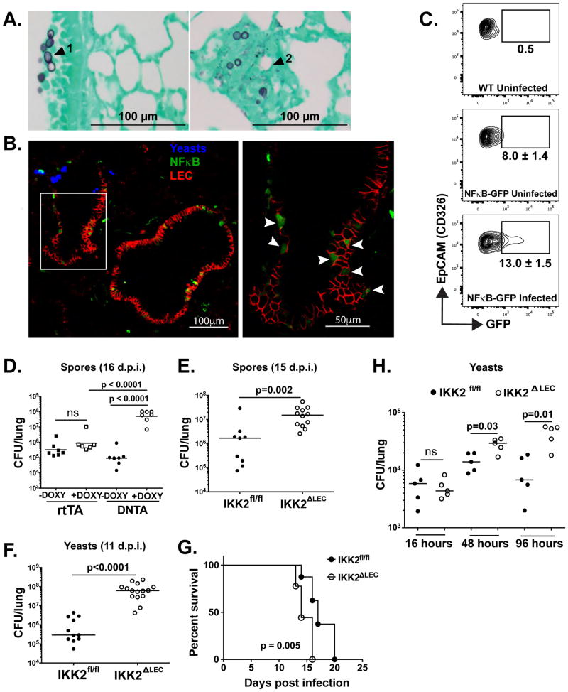
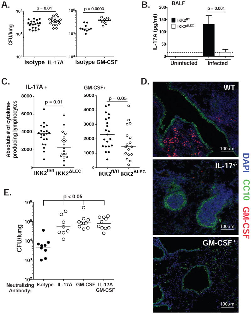
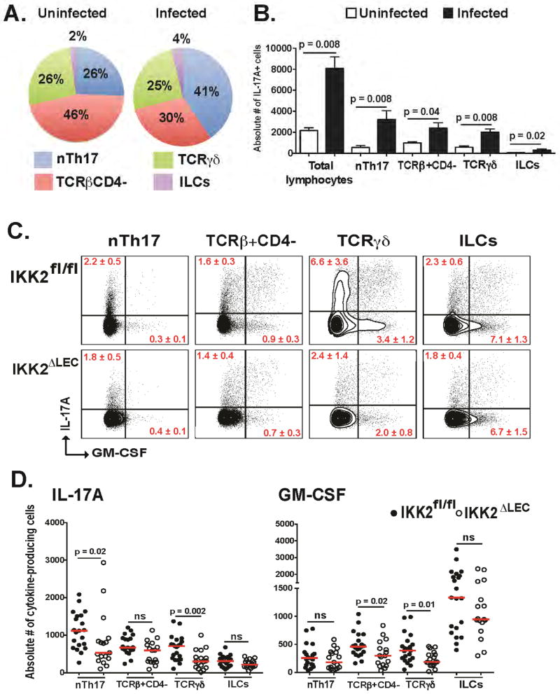
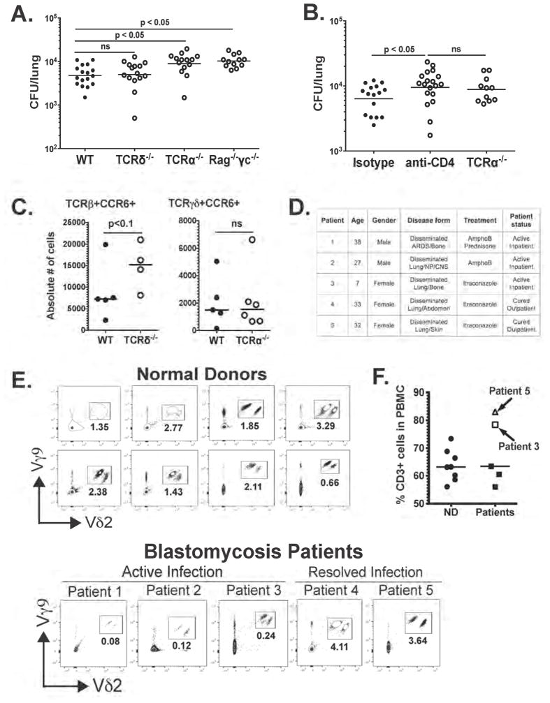
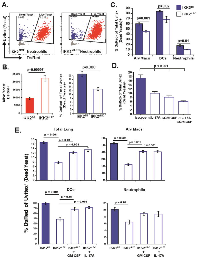
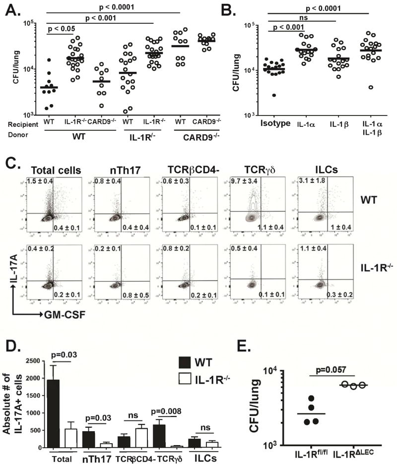
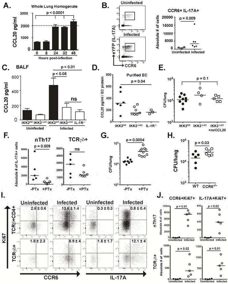
Lung epithelial cells (LECs) are strategically positioned in the airway mucosa to provide barrier defense. LECs also express pattern recognition receptors and a myriad of immune genes, but their role in immunity is often concealed by the activities of "professional" immune cells, particularly in the context of fungal infection. Here, we demonstrate that NF-κB signaling in LECs is essential for immunity against the pulmonary fungal pathogen Blastomyces dermatitidis. LECs orchestrate innate antifungal immunity by augmenting the numbers of interleukin-17A (IL-17A)- and granulocyte-macrophage colony-stimulating factor (GM-CSF)-producing innate lymphocytes, specifically "natural" Th17 (nTh17) cells. Innate lymphocyte-derived IL-17A and GM-CSF in turn enable phagocyte-driven fungal killing. LECs regulate the numbers of nTh17 cells via the production of chemokines such as CCL20, a process dependent on IL-1α-IL-1 receptor (IL-1R) signaling on LECs. Therefore, LECs orchestrate IL-17A- and GM-CSF-mediated immunity in an IL-1R-dependent manner and represent an essential component of innate immunity to pulmonary fungal pathogens.

Keywords: epithelial cells; fungi; innate immunity; lymphocytes.


Conflict of interest statement

Declaration of Interests. The authors declare no competing interests.

Figures

Figure 1
Figure 1. LEC mount a robust, NFκB-dependent antifungal response early upon infection (see also Fig. S1)
(A) WT mice were infected i.t. with 2×10⁵ yeasts and 24 h.p.i. lungs were inflated and fixed in formalin. Tissue sections are stained with gomori methanimine silver. (B) NFκB-GFP reporter mice were infected with 5×10⁵ yeast labeled with calcofluor and cryosections of lung tissue were analyzed by confocal microscopy ~3.5 h.p.i. Blue denotes yeast, green GFP (NFκB), and red CD326+ cells (LEC). Left photo = 20X mag. The right photo is the area denoted in white box at 60X mag. (C) NFκB-GFP reporter mice were infected as in panel B and analyzed for GFP expression in Ep-CAM+ (CD326+) cells from lungs at 24 h.p.i. Gates depict mean±SD % GFP+ cells (uninfected group n=5, infected group n=4). (D–F) DNTA or IKK2ΔLEC mice and littermate controls were infected i.t. with 1×10⁵ spores or 2×10⁴ yeast and lung CFU quantified at indicated times. Each symbol denotes a mouse. One-Way ANOVA with Bonferroni’s correction (D) and Mann-Whitney test (E–F). (G) IKK2fl/fl and IKK2ΔLEC were infected with yeast and monitored for survival over 20 days. Log-rank test. (H) IKK2fl/fl and IKK2ΔLEC were infected and analyzed for CFU as indicated in (D–F). Mann-Whitney test.
Figure 2
Figure 2. LEC-mediated antifungal immunity is IL-17A- and GM-CSF-dependent (see also Fig. S2)
(A) WT mice were infected i.t. with yeast. IL-17A and GM-CSF were neutralized as noted in Methods. At 48 h.p.i., lung CFU was enumerated. Each panel represents a separate experiment (2 pooled experiments depicted in left panel); each symbol denotes one mouse. Mann-Whitney test (48 h.p.i.). (B) IKK2fl/fl and IKK2ΔLEC mice were infected i.t. with yeast and IL-17A content was analyzed by ELISA in BALF at 48 h.p.i. A representative of 4 experiments is depicted. Mann-Whitney test. (C) IKK2fl/fl and IKK2ΔLEC mice were infected i.t. with yeast. At 48 h.p.i., lung cell suspensions were analyzed by flow cytometry for intracellular IL-17A and GM-CSF in lymphocytes (CD90.2+ CD44hi). Two pooled experiments depicted. Mann-Whitney test. (D) WT, IL-17A−/− and GM-CSF−/− were infected with 5×10⁵ yeasts; at 48 h.p.i., lungs were analyzed by confocal imaging. Club cells depicted in green (CC10), nuclei in blue (DAPI) and GM-CSF in red. (E) Procedure was done as in A with one further injection of antibody at 48 h.p.i. At 96 h.p.i., lung CFU were quantified. Data representative of 3 experiments. Kruskal-Wallis with Dunn’s multiple comparison.
Figure 3
Figure 3. Ablation of NFκB in LEC results in decreased numbers of IL-17A- and GM-CSF-producing innate lymphocytes (see also Fig. S3)
(A–B) IL-17creRosa26ReYFP mice were infected i.t. with yeast. Lung cell suspensions were analyzed by flow cytometry at 48 h.p.i. to identify IL-17A+ innate lymphocytes. Cells were gated as CD90.2+IL-17+ and the proportion (A) and number (B) of nTh17, TCRβ+ CD4, TCRγδ, and ILCs quantified. Total lymphocytes (B) means IL-17+ cells within the total CD90.2+ population. A representative of 3 experiments depicted; n=5/group. Mann-Whitney test (B). (C–D) IKK2fl/fl and IKK2ΔLEC mice were infected i.t. with yeast and the proportion (C) and number (D) of indicated lymphocyte populations quantified by flow cytometry at 48 h.p.i. The TCRβ+CD4 gate includes CD8+ T cells. Concatenated plots depicted in C (n=11 in IKK2fl/fl, n=9 in IKK2ΔLEC). Two pooled experiments depicted in D; each symbol is one mouse. Mann-Whitney test.
Figure 4
Figure 4. nTh17 cells are indispensable for innate antifungal immunity (see also Fig. S4)
(A) WT, TCRδ−/− TCRα−/− and Rag−/−γc−/− mice were infected i.t. with yeast and at 48 h.p.i. lung CFU was enumerated. Two pooled experiments depicted; each symbol denotes one mouse. One-Way ANOVA with Bonferroni’s correction. (B) WT (isotype), CD4-neutralized, and TCRα−/− mice were infected i.t. with yeast and CFU counted at 48 h.p.i. α-CD4 given i.v. at the time of infection. Two pooled experiments depicted; each symbol denotes a mouse. One-way ANOVA with Bonferroni’s correction. (C) WT, TCRδ−/− TCRα−/− mice were infected i.t. with yeast. At 48 h.p.i., the number of CCR6+ cells among TCRβ+ and TCRγδ+ cells was quantified by flow cytometry. Experiment is representative of two performed. Mann-Whitney test. (D) Summary of blastomycosis patient features. (E) Frequency of Vγ9+Vδ2+ among total CD3+ cells in peripheral blood of healthy donors and patients with active or resolved blastomycosis was quantified by flow cytometry. Each dot plot depicts a donor. (F) Proportion of total CD3+ cells in normal donors (ND) and patients with blastomycosis.
Figure 5
Figure 5. Collaborative killing of yeast by alveolar MØ, DCs, and neutrophils is dependent on LEC NFκB, IL-17A and GM-CSF
(A–B) IKK2fl/fl and IKK2ΔLEC mice were infected i.t. with DsRed yeast stained with Uvitex. The proportion and number of live (red) and dead (blue) yeast among total yeasts (red+blue) were quantified by flow cytometry. Two pooled experiments depicted. (C) Proportion of yeast-associated (uvitex+) alveolar MØ (CD11c+Siglec F+), DCs (Siglec F CD11c+ MHCII+) and neutrophils (Siglec F CD11b+Ly6G+) that were DsRed- (associated with dead yeast). Two pooled experiments depicted. (D) WT mice were infected i.t. with DsRed yeast and the proportion of dead yeasts was calculated by flow cytometry. IL-17A and GM-CSF were neutralized as noted in Methods. Data representative of 3 experiments. One-way ANOVA with Tukey’s multiple comparison. (E) IKK2fl/fl and IKK2ΔLEC were infected with DsRed yeast. rIL-17A and rGM-CSF were given i.t. together with the inoculum. 48 h.p.i lung cell suspensions were analyzed by flow cytometry to quantify the proportion of dead yeast (DeRedUvitex+) in total lung homogenate, and proportoin of phagocytes associated with dead yeast. One-Way ANOVA with Tukey’s multiple comparison.
Figure 6
Figure 6. LEC-mediated antifungal immunity is dependent on the IL-1α/IL-1R signaling axis (see also Fig. S5 and S6)
(A) Bone marrow chimeric mice were infected i.t. with yeast and lung CFU counted at 48 h.p.i. A representative of 2 experiments is shown. One-way ANOVA with Bonferroni’s correction. (B) WT mice were infected i.t. with yeast and lung CFU counted at 48 h.p.i. IL-1α and IL-1β were neutralized at the time of infection. Two pooled experiments depicted. One-way ANOVA with Bonferroni’s correction. (C–D) IL-17A and GM-CSF production by innate lymphocytes in WT and IL-1R−/− mice were analyzed by intracellular cytokine staining. Proportion (C) and number (D) of cytokine-producing cells are depicted. Concatenated plots depicted in C (n=5 WT and 5 IL-1R−/−). A representative experiment of two is shown. Mann Whitney test. (E) IL-1Rfl/fl and IL-1RΔLEC mice were infected with yeast and CFU counted at 48 h.p.i. Mann Whitney test.
Figure 7
Figure 7. IL-1R signaling regulates CCL20 production by LEC and CCL20 partners with other chemokines to regulate nTh17 numbers (see also Fig. S7)
(A) CCL20 levels in lung homogenate were quantified by ELISA at various times after infection with yeast. One-way ANOVA with Bonferroni’s correction. Dotted line is upper limit of detection (B) Proportion (left) and number (right) of CCR6+IL-17A+ nTh17 cells in IL-17creRosa 26ReYFP mice after infection. Mann-Whitney test. (C) CCL20 levels in BALF of IKK2fl/fl, IKK2ΔLEC and IL-1R−/− quantified by ELISA 48 h.p.i. Four pooled experiments depicted. One-way ANOVA with Bonferroni’s correction. (D) LEC (CD31CD45CD326+) were purified from infected mice (48 h.p.i.) and levels of CCL20 in cell lysates quantified by ELISA. Two pooled experiments depicted. One-way ANOVA. (E) Yeasts were given i.t. alone or together with rCCL20. At 48 h.p.i., lung CFU was quantified. One-way ANOVA. (F) IL-17creRosa26ReYFP were infected with yeast i.t. alone or together with PTx. At 48h.p.i., lungs were harvested and the number of IL-17A+ nTh17 and TCRγδ+ cells quantified by flow cytometry. Mann-Whitney test. (G) Yeasts were given i.t. alone or together with PTx. At 48 h.p.i, lung CFU was quantified. Mann-Whitney test. (H) Mice were infected with yeast i.t. and at 48 h.p.i. lung CFU was quantified. One-tailed, unpaired t test. (I–J) WT mice were infected with yeast and after 48h the proportion (H) and number (I) of CCR6+Ki67+ and IL-17A+ Ki67+ cells were quantified by flow cytometry. Ki67+IL-17A+ and Ki67+CCR6+ cells in the TCRβ+CD4+ gate (H) denote proliferating nTh17 cells (enumerated in I). Mann-Whitney test (I).

References

    1. Aimanianda V, Bayry J, Bozza S, Kniemeyer O, Perruccio K, Elluru SR, Clavaud C, Paris S, Brakhage AA, Kaveri SV, et al. Surface hydrophobin prevents immune recognition of airborne fungal spores. Nature. 2009;460:1117–1121. - PubMed
    1. Balbi B, Valle MT, Oddera S, Giunti D, Manca F, Rossi GA, Allegra L. T-lymphocytes with gamma delta+ V delta 2+ antigen receptors are present in increased proportions in a fraction of patients with tuberculosis or with sarcoidosis. The American review of respiratory disease. 1993;148:1685–1690. - PubMed
    1. Bar E, Whitney PG, Moor K, Reis ESC, Leibundgut-Landmann S. IL-17 Regulates Systemic Fungal Immunity by Controlling the Functional Competence of NK Cells. Immunity 2014 - PubMed
    1. Bello-Irizarry SN, Wang J, Olsen K, Gigliotti F, Wright TW. The alveolar epithelial cell chemokine response to pneumocystis requires adaptor molecule MyD88 and interleukin-1 receptor but not toll-like receptor 2 or 4. Infection and immunity. 2012;80:3912–3920. - PMC - PubMed
    1. Carding SR, Egan PJ. Gammadelta T cells: functional plasticity and heterogeneity. Nature reviews Immunology. 2002;2:336–345. - PubMed

Publication types

Substances

LinkOut - more resources