. 2018 Jan 16:2018:4720532.
doi: 10.1155/2018/4720532. eCollection 2018.

Palmitoylethanolamide Dampens Reactive Astrogliosis and Improves Neuronal Trophic Support in a Triple Transgenic Model of Alzheimer's Disease: In Vitro and In Vivo Evidence

Affiliations

Palmitoylethanolamide Dampens Reactive Astrogliosis and Improves Neuronal Trophic Support in a Triple Transgenic Model of Alzheimer's Disease: In Vitro and In Vivo Evidence

Maria Rosanna Bronzuoli et al. Oxid Med Cell Longev. .

Abstract

Alzheimer's disease (AD) is a neurodegenerative disorder responsible for the majority of dementia cases in elderly people. It is widely accepted that the main hallmarks of AD are not only senile plaques and neurofibrillary tangles but also reactive astrogliosis, which often precedes detrimental deposits and neuronal atrophy. Such phenomenon facilitates the regeneration of neural networks; however, under some circumstances, like in AD, reactive astrogliosis is detrimental, depriving neurons of the homeostatic support, thus contributing to neuronal loss. We investigated the presence of reactive astrogliosis in 3×Tg-AD mice and the effects of palmitoylethanolamide (PEA), a well-documented anti-inflammatory molecule, by in vitro and in vivo studies. In vitro results revealed a basal reactive state in primary cortical 3×Tg-AD-derived astrocytes and the ability of PEA to counteract such phenomenon and improve viability of 3×Tg-AD-derived neurons. In vivo observations, performed using ultramicronized- (um-) PEA, a formulation endowed with best bioavailability, confirmed the efficacy of this compound. Moreover, the schedule of treatment, mimicking the clinic use (chronic daily administration), revealed its beneficial pharmacological properties in dampening reactive astrogliosis and promoting the glial neurosupportive function. Collectively, our results encourage further investigation on PEA effects, suggesting it as an alternative or adjunct treatment approach for innovative AD therapy.


Figures

Figure 1
Figure 1
Study designs. Schematic representation of the experimental designs in (a) primary 3×Tg-AD and non-Tg astrocytes, (b) primary 3×Tg-AD neurons, and (c) in vivo experiments in 3×Tg-AD and non-Tg mice.
Figure 2
Figure 2
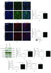
Study of parameters related to reactive astrogliosis in 3×Tg-AD and non-Tg primary astrocytes. (a) Representative fluorescent photomicrographs of GFAP (green) and (b) signal quantification in both non-Tg (white bar) and 3×Tg-AD (black bar) primary astrocytes. (c) Representative fluorescent photomicrographs of S100B (red) and (d) signal quantification in both non-Tg (white bar) and 3×Tg-AD (black bar) primary astrocytes. Nuclei were stained with DAPI (blue). Scale bar is 50 μm. Fluorescence analysis is expressed as ΔF/F0. (e) Representative bands and Western blot densitometric analysis of (f) GFAP, (g) S100B, (h) iNOS, and (i) COX-2. β-Actin was used as loading control. Results are expressed as percentage of the mean control value (non-Tg cells). Experiments were performed three times in triplicate. Data are presented as mean ± SEM. The statistical analysis was performed by Student's t-test (P < 0.05 and ∗∗P < 0.01 versus non-Tg group).
Figure 3
Figure 3
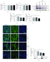
Effect of PEA treatment on astrocyte and neuronal viability and reactive astrogliosis in 3×Tg-AD cells. Evaluation of (a) astrocyte and (b) neuronal viability tested by neutral red uptake assay after 24 h PEA treatment (0.01–0.1–1 μM). (c) Representative immunoreactive signals and Western blot densitometric analysis of (d) GFAP and (e) iNOS. β-Actin was used as loading control. Results are expressed as percentage of the mean control value (CTRL). (f) Representative fluorescent photomicrographs of GFAP (green) staining in 3×Tg-AD primary astrocytes. Nuclei were stained with DAPI (blue). Scale bar is 50 μm. (g) Fluorescence analysis is expressed as ΔF/F0. Experiments were performed three times in triplicate. Data are presented as mean ± SEM. The statistical analysis was performed by one-way ANOVA followed by Bonferroni's post hoc multiple comparison test (P < 0.05 and ∗∗∗P < 0.001 versus CTRL group).
Figure 4
Figure 4
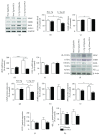
Effect of chronic um-PEA on reactive astrogliosis and Aβ(1–42) expression in the FC of 3×Tg-AD and non-Tg mice. (a) Representative bands from RT-PCR performed in FC homogenates for GFAP, S100B, iNOS, and COX-2, and (b–e) densitometric analysis of the corresponding signals normalized to GAPDH. (f) Representative immunoreactive species and Western blot densitometric analysis of (g) GFAP, (h) S100B, (i) COX-2, (j) iNOS, and (k) Aβ(1–42). β-Actin was used as loading control. Results are expressed as percentage of the mean control value (non-Tg/placebo). Experiments were performed three times in triplicate. Data are presented as mean ± SEM. The statistical analysis was performed by two-way ANOVA followed by Bonferroni's post hoc multiple comparison test (P < 0.05 and ∗∗∗P < 0.001 versus non-Tg/placebo group; °P < 0.05, °°P < 0.01, and °°°P < 0.001 versus 3×Tg-AD/placebo group).
Figure 5
Figure 5
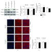
Effect of chronic um-PEA on neuronal support and survival in FC of 3×Tg-AD and non-Tg mice. (a) Representative immunoreactive species and Western blot densitometric analysis of (b) BDNF and (c) MAP2. β-Actin was used as loading control. Results are expressed as percentage of the mean control value (non-Tg/placebo). (d) Representative fluorescent photomicrographs of MAP2 (red) staining in FC of 6-month-old non-Tg and 3×Tg-AD mice, placebo- or um-PEA-treated. Nuclei were stained with DAPI (blue). Scale bar is 50 μm. (e) Fluorescence analysis is expressed as ΔF/F0. Experiments were performed three times in triplicate. Data are presented as mean ± SEM. The statistical analysis was performed by two-way ANOVA followed by Bonferroni's post hoc multiple comparison test (P < 0.05 versus non-Tg/placebo group; °P < 0.05 versus 3×Tg-AD/placebo group).

References

    1. Anand R., Gill K. D., Mahdi A. A. Therapeutics of Alzheimer’s disease: past, present and future. Neuropharmacology. 2014;76:27–50. doi: 10.1016/j.neuropharm.2013.07.004. - DOI - PubMed
    1. Braak H., Braak E. Neuropil threads occur in dendrites of tangle—bearing nerve cells. Neuropathology and Applied Neurobiology. 1988;14(1):39–44. doi: 10.1111/j.1365-2990.1988.tb00864.x. - DOI - PubMed
    1. Merz P., Wisniewski H., Somerville R., Bobin S. A., Masters C. L., Iqbal K. Ultrastructural morphology of amyloid fibrils from neuritic and amyloid plaques. Acta Neuropathology. 1983;60(1-2):113–124. doi: 10.1007/BF00685355. - DOI - PubMed
    1. Rodríguez J. J., Olabarria M., Chvatal A., Verkhratsky A. Astroglia in dementia and Alzheimer’s disease. Cell Death & Differentiation. 2009;16(3):378–385. doi: 10.1038/cdd.2008.172. - DOI - PubMed
    1. Akiyama H., Barger S., Barnum S., Bradt B., Bauer J., Cole G. M. Inflammation and Alzheimer’s disease. Neurobiology of Aging. 2000;21(3):383–421. doi: 10.1016/S0197-4580(00)00124-X. - DOI - PMC - PubMed