

A new worm infiltrating the human cornea: A report of three cases
- PMID: 29577104
- PMCID: PMC5861503
- DOI: 10.1016/j.ajoc.2018.01.013
A new worm infiltrating the human cornea: A report of three cases
Abstract
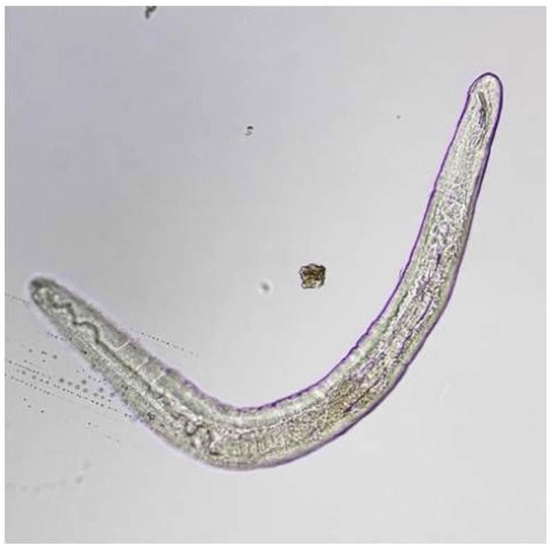
Purpose: To characterize a new species of parasitic nematode that triggers uveitis.
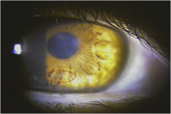
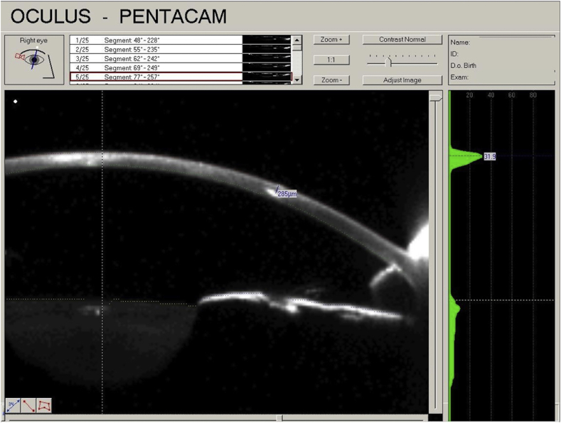
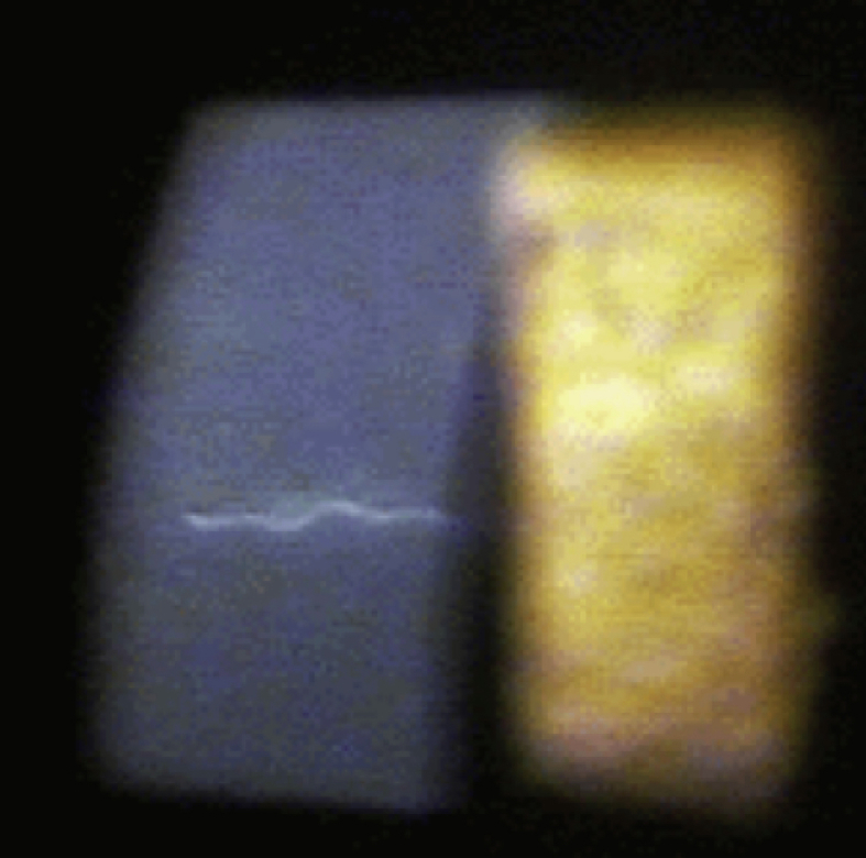
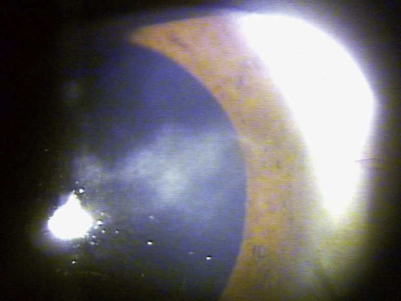
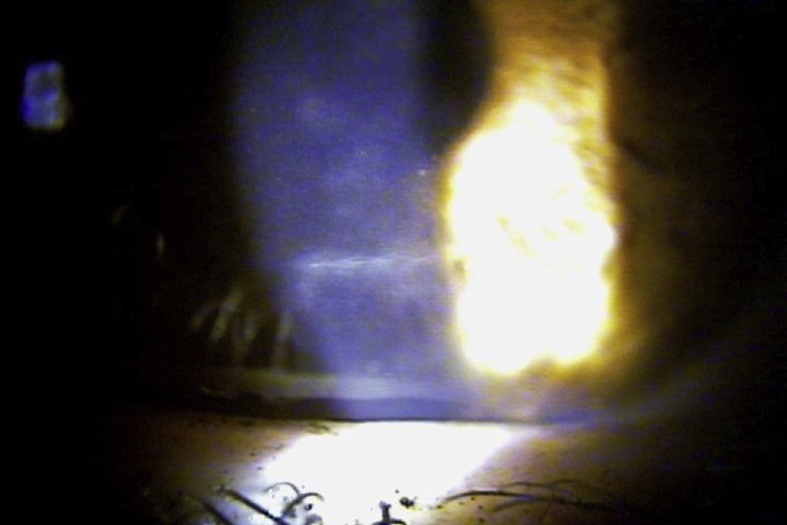
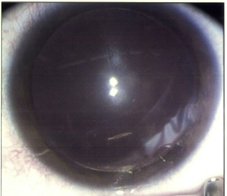
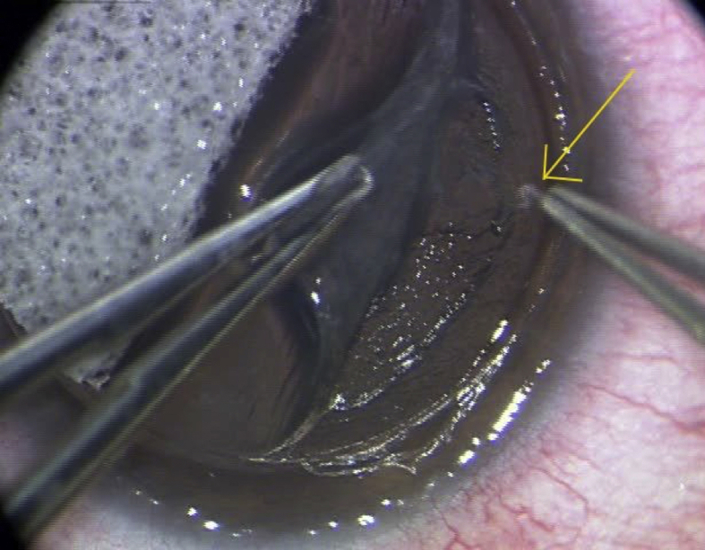
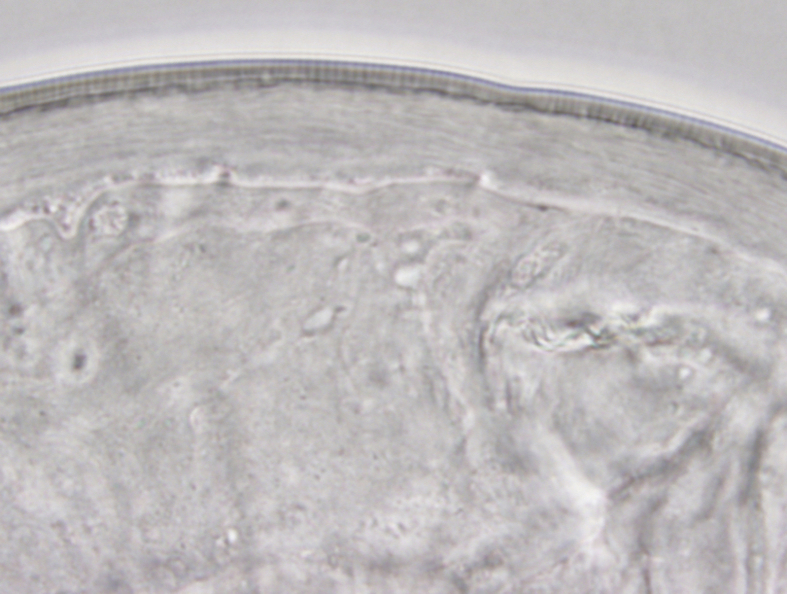
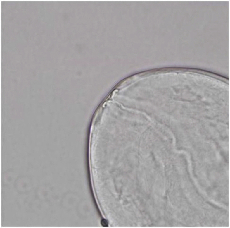
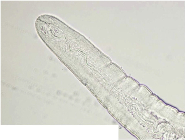
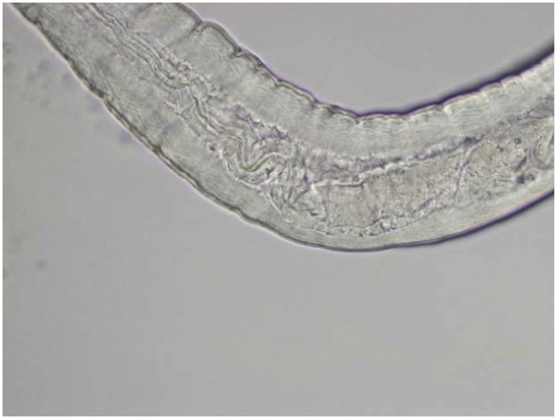
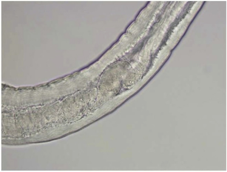
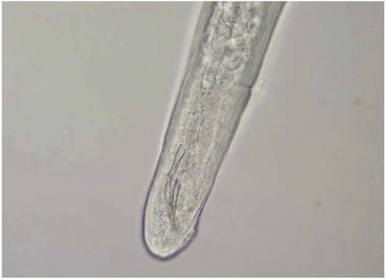
Observations: Three previously healthy, relatively young people each contracted a corneal stromal nematode that, upon surgical removal and examination, did not match any known nematodes. Clinical ocular findings included corneal opacification, visible corneal worms, conjunctival injection, and uveitis.
Conclusions and importance: The three cases presented here represent a previously undescribed parasitic infection of the cornea by an unidentified nematode. These findings may represent a previously unrecognized zoonotic infection from wildlife sources and potentially a newly documented nematode requiring description. Future clinical findings regarding this newly described nematode are needed to further develop our understanding of the disease.
Keywords: Cornea; Nematode; Ocular; Parasite; Stroma; Uveitis.
Figures
Similar articles
-
Zoonotic Onchocerca (Nematoda:Filarioidea) in the cornea of a Colorado resident.Ophthalmology. 1998 Aug;105(8):1494-7. doi: 10.1016/S0161-6420(98)98035-6. Ophthalmology. 1998. PMID: 9709764
-
Immune mechanisms in Onchocerca volvulus-mediated corneal disease (river blindness).Parasite Immunol. 2000 Dec;22(12):625-31. doi: 10.1046/j.1365-3024.2000.00345.x. Parasite Immunol. 2000. PMID: 11123754 Review.
-
A combined parasitological molecular approach for noninvasive characterization of parasitic nematode communities in wild hosts.Mol Ecol Resour. 2015 Sep;15(5):1112-9. doi: 10.1111/1755-0998.12382. Epub 2015 Feb 26. Mol Ecol Resour. 2015. PMID: 25644900
-
Limbal stem cell transplantation: an evidence-based analysis.Ont Health Technol Assess Ser. 2008;8(7):1-58. Epub 2008 Oct 1. Ont Health Technol Assess Ser. 2008. PMID: 23074512 Free PMC article.
-
Elucidating the molecular and developmental biology of parasitic nematodes: Moving to a multiomics paradigm.Adv Parasitol. 2020;108:175-229. doi: 10.1016/bs.apar.2019.12.005. Epub 2020 Jan 31. Adv Parasitol. 2020. PMID: 32291085 Review.
Cited by
-
Case report: an unusual unilateral pterygium - a secondary pterygium caused by parasitosis in the scleral fistula.BMC Ophthalmol. 2021 Sep 6;21(1):323. doi: 10.1186/s12886-021-02083-2. BMC Ophthalmol. 2021. PMID: 34488674 Free PMC article.
-
Concomitant Potentially Contagious Factors Detected in Poland and Regarding Acanthamoeba Strains, Etiological Agents of Keratitis in Humans.Microorganisms. 2024 Nov 28;12(12):2445. doi: 10.3390/microorganisms12122445. Microorganisms. 2024. PMID: 39770648 Free PMC article.
-
New insights into the clinico-histopathological and molecular features of Pelecitus (Filarioidea: Onchocercidae) from a raptor bird.Parasitol Res. 2018 Oct;117(10):3319-3325. doi: 10.1007/s00436-018-6009-1. Epub 2018 Jul 13. Parasitol Res. 2018. PMID: 30006807
References
-
- Magill A.J., Strickland G.T., Maguire J.H., Ryan E.T., Solomon T. Elsevier; China: 2000. Helminthic Infections. Hunter's Tropical Medicine and Emerging Infectious Disease.
-
- Streilein J. Karger; Basel, Switzerland: 1999. Regional Immunity and Ocular Immune Privilege. Immune Response and the Eye. - PubMed
Publication types
LinkOut - more resources
Full Text Sources
Other Literature Sources
