Review
. 2018 Jan 1;9(1):235-248.
doi: 10.1080/19491034.2018.1456217.

Lipodystrophic syndromes due to LMNA mutations: recent developments on biomolecular aspects, pathophysiological hypotheses and therapeutic perspectives

Affiliations
Review

Lipodystrophic syndromes due to LMNA mutations: recent developments on biomolecular aspects, pathophysiological hypotheses and therapeutic perspectives

Corinne Vigouroux et al. Nucleus. .

Abstract

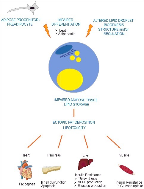
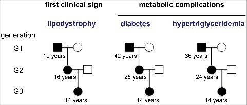
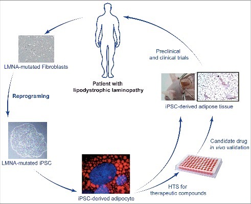
Mutations in LMNA, encoding A-type lamins, are responsible for laminopathies including muscular dystrophies, lipodystrophies, and premature ageing syndromes. LMNA mutations have been shown to alter nuclear structure and stiffness, binding to partners at the nuclear envelope or within the nucleoplasm, gene expression and/or prelamin A maturation. LMNA-associated lipodystrophic features, combining generalized or partial fat atrophy and metabolic alterations associated with insulin resistance, could result from altered adipocyte differentiation or from altered fat structure. Recent studies shed some light on how pathogenic A-type lamin variants could trigger lipodystrophy, metabolic complications, and precocious cardiovascular events. Alterations in adipose tissue extracellular matrix and TGF-beta signaling could initiate metabolic inflexibility. Premature senescence of vascular cells could contribute to cardiovascular complications. In affected families, metabolic alterations occur at an earlier age across generations, which could result from epigenetic deregulation induced by LMNA mutations. Novel cellular models recapitulating adipogenic developmental pathways provide scalable tools for disease modeling and therapeutic screening.

Keywords: Lamin A/C; adipose tissue; anticipation; differentiation; epigenetics; extracellular matrix; induced pluripotent stem cells; lipodystrophy; metreleptin; senescence.


Figures

Figure 1.
Figure 1.
Impaired adipose tissue lipid storage induces metabolic complications in lipodystrophic syndromes. Most genes involved in lipodystrophic syndromes have been shown to regulate adipocyte differentiation, triglycerides synthesis, lipolysis, and/or lipid droplet structure or biogenesis. Impaired storage of excess energy as triglycerides in adipocytes leads to ectopic fat deposition and lipotoxicity in several tissues such as muscle, heart, liver and pancreas, resulting in post-receptor insulin resistance, dyslipidemia and liver steatosis.
Figure 2.
Figure 2.
An early detrimental remodeling of adipose tissue extracellular matrix could contribute to the pathophysiology of LMNA-associated lipodystrophies. Extracellular matrix alterations induced by lipodystrophy-causing LMNA mutations could hamper adipocyte differentiation and limit the expandability of adipose tissue, triggering adipocyte dysfunction and metabolic defects.
Figure 3.
Figure 3.
Several laminopathies are characterized by tissular fibrosis, increased TGF-beta signaling and/or metalloproteinase expression/activity. Extracellular matrix alterations at the level of adipose tissue, vascular wall or heart have been described in several laminopathies and participate to the clinical phenotype and to the complications of laminopathies. ,
Figure 4.
Figure 4.
Vascular effects of LMNA mutations causing lipodystrophy. A. Prelamin-A physiologically undergoes a complex post-translational maturation process affecting its C-terminal CaaX motif. After farnesylation of the carboxy-terminal cysteine, aaX amino acids are removed, farnesyl-cysteine is carboxymethylated, then the 15 C-terminal amino acids are cleaved by the metalloprotease ZMPSTE24 to produce mature lamin A. B. Lipodystrophy-causing mutations in LMNA lead to accumulation of farnesylated prelamin A and nuclear envelope disorganization. C. Accumulation of prelamin A pathogenic variants at the nuclear envelope induces oxidative stress, inflammation and cellular senescence. These cellular alterations contribute to endothelial cell dysfunction and to osteoblastic transdifferentiation of vascular smooth muscle cell, promoting atherosclerosis and vascular calcification.
Figure 5.
Figure 5.
Anticipation of metabolic complications in lipodystrophic laminopathies. The comparison of the natural history and disease severity in familial forms of lipodystrophic syndromes due to LMNA pathogenic variants reveals similar characteristics and age at onset for lipodystrophy, but an anticipation of metabolic complications over generations. The mean age at onset of each clinical sign is indicated below symbols representing patients from different generations.
Figure 6.
Figure 6.
A new cellular tool for studying adipocyte development in vitro. Patients’ primary cells reprogramming into induced Pluripotent Stem Cells (iPSCs) allow to study several relevant cells types that would be otherwise inaccessible. This also avoids using non-physiological lamin overexpression strategies. Differentiation of iPS cells originating from patients with lipodystrophic laminopathies into adipocytes through a developmentally relevant protocol also provides an unlimited source of cells for high throughput screening (HTS) of therapeutic compounds, opening perspectives for the treatment of these rare diseases.

References

    1. Cuthbertson DJ, Steele T, Wilding JP, Halford JC, Harrold JA, Hamer M, Karpe F. What have human experimental overfeeding studies taught us about adipose tissue expansion and susceptibility to obesity and metabolic complications? Int J Obes (Lond). 2017;41(6):853–865. - PubMed
    1. Stefan N, Schick F, Haring HU. Causes, Characteristics, and Consequences of Metabolically Unhealthy Normal Weight in Humans. Cell Metab. 2017;26(2):292–300. - PubMed
    1. Pellegrinelli V, Carobbio S, Vidal-Puig A. Adipose tissue plasticity: how fat depots respond differently to pathophysiological cues. Diabetologia. 2016;59(6):1075–88. - PMC - PubMed
    1. Lotta LA, Gulati P, Day FR, Payne F, Ongen H, van de Bunt M, Gaulton KJ, Eicher JD, Sharp SJ, Luan J, et al. . Integrative genomic analysis implicates limited peripheral adipose storage capacity in the pathogenesis of human insulin resistance. Nat Genet. 2017;49(1):17–26. - PMC - PubMed
    1. Barroso I, Gurnell M, Crowley VE, Agostini M, Schwabe JW, Soos MA, Maslen GL, Williams TD, Lewis H, Schafer AJ, et al. . Dominant negative mutations in human PPARgamma associated with severe insulin resistance, diabetes mellitus and hypertension. Nature. 1999;402(6764):880–3. - PubMed

Publication types

LinkOut - more resources