Review
. 2018 May;40(5):e1700249.
doi: 10.1002/bies.201700249. Epub 2018 Mar 26.

Multi-Invasion-Induced Rearrangements as a Pathway for Physiological and Pathological Recombination

Affiliations
Review

Multi-Invasion-Induced Rearrangements as a Pathway for Physiological and Pathological Recombination

Aurèle Piazza et al. Bioessays. 2018 May.

Abstract

Cells mitigate the detrimental consequences of DNA damage on genome stability by attempting high fidelity repair. Homologous recombination templates DNA double-strand break (DSB) repair on an identical or near identical donor sequence in a process that can in principle access the entire genome. Other physiological processes, such as homolog recognition and pairing during meiosis, also harness the HR machinery using programmed DSBs to physically link homologs and generate crossovers. A consequence of the homology search process by a long nucleoprotein filament is the formation of multi-invasions (MI), a joint molecule in which the damaged ssDNA has invaded more than one donor molecule. Processing of MI joint molecules can compromise the integrity of both donor sites and lead to their rearrangement. Here, two mechanisms for the generation of rearrangements as a pathological consequence of MI processing are detailed and the potential relevance for non-allelic homologous recombination discussed. Finally, it is proposed that MI-induced crossover formation may be a feature of physiological recombination.

Keywords: copy number variation; genomic instability; homologous recombination; non-allelic homologous recombination; repeated sequence; structural variant; translocation.


Conflict of interest statement

Conflict of Interest

The authors declare no conflict of interest.

Figures

Figure 1.
Figure 1.
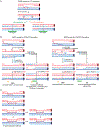
Outline of multi-invasion-induced rearrangements and its regulation by cis and trans-acting factors.
Figure 2.
Figure 2.
MIR1 mechanism. A) Top and bottom strands processing results in a chromosomal translocation. Formation of each translocated DNA strand can be independent from each other. The only kinetic constraint of MIR1 is the cleavage of I5 prior to being reached by the synthesis initiated at T5. MIR1 leaves as immediate products: 1) the original DSB, which has lost its 3′ extremity involved in MIR and that can be repaired by HR using the homolog as a template; 2) the translocation chromatid; and 3) and 4) two oriented single-ended DSBs (the left side of the T donor with a 3′ protruding extremity and the right side of the I invasion with a 5′ protruding extremity). Both can be repaired with or without additional rearrangements, depending on their sequence context. The question mark indicates uncertainties regarding the nature and position of the cleavage events at I5. B) Outline of MIR1 initiated by a ssDNA gap. Dashed lines indicate recombination-associated DNA synthesis.
Figure 3.
Figure 3.
MIR2 mechanism. The internal and distal donors must share downstream sequence homology (depicted in green). The endonucleolytic processing of the I invasion is identical to MIR1. The question mark indicates uncertainties regarding the nature and position of the cleavage events at I5. MIR2 produces a single, 5′ protruding one-ended DSB which upon resection is annealed to the displaced extended invading strand at the T donor. MIR2 leaves as immediate products: 1) the original DSB, which has lost its 3′ extremity involved in MIR and can be repaired by HR using the homolog as a template; 2) an apparent translocation which formally is an insertion. The chromosome with the terminal (red) donor is intact. Hence, MIR2 does not propagate additional DSB. Dashed lines indicate recombination-associated DNA synthesis.
Figure 4.
Figure 4.
MIR-based mechanisms for CO and mcJMs formation. A) The DSBR model for CO formation. Colored arrows indicate the two possible orientations for dHJ cleavage resulting in a CO. Dotted strands result from DNA synthesis. The green bars represent the CO region, which in both cases encompass the DSB site (dotted black line). B) Examples of MIR1- (left) and MIR2-based (right) mechanisms for NCO and CO formation upon inter-sister MI. The two initial DSB ends (a and b) and the two DSB ends generated upon MIR (c and d) are labeled. The particular outcome of their repair depends (i) on the relative timing of formation and processing of the “c” and “d” DSBs, and (ii) the specific template and repair pathway employed. Additional examples of inter-homolog events and the various repair outcomes of downstream DSBs are depicted in Figure S1, Supporting Information. Orange bars denote gapped regions prone to gene conversion. Dotted strands result from DNA synthesis. The green bars represent the CO regions, which never encompass the DSB site (dotted black line). The outcome description follows the nomenclature of Ref. [21]. C) Example of tri-parental mcJMs formation from an inter-homolog MI precursor. The template switches can readily occur upon DNA strand exchange junctions migration towards the DNA synthesis direction. All strands are in the parental configurations. Dotted strands result from DNA synthesis. Double-headed arrows indicate equivalence of the intermediates.
Figure 5.
Figure 5.
MIR-based mechanisms for NAHR. A) DSBR model for NAHR. The DSB must occur within a repeat. * indicates homeologous invasions. B) Example of a MIR1-induced inter-chromatidal deletion event. In this scenario, the only homeologous invasion occurs internally, the distal invasion being allelic. The repair of the secondary DSBs will lead either to a loss of the intervening sequence (left) or a reciprocal duplication (right). The translocation produced by MIR is boxed in red. C) Other possible inter-chromatidal NAHR events depending on the repeats invaded and the pathway employed to repair the secondary DSBs. The translocations produced by MIR are boxed in red. For each outcome, a detailed mechanism is presented in Figure S2, Supporting Information. D) Schematic representation of the distribution around repeated regions of DSB sites prone to induce NAHR events according to the DSBR (top) and the MIR (bottom) models. For simplicity, heteroduplex regions are not drawn in this figure.

References

    1. Bell JC, Kowalczykowski SC, Annu. Rev. Biochem 2016, 85, 193. - PubMed
    1. Forget AL, Kowalczykowski SC, Nature 2012, 482, 423. - PMC - PubMed
    1. Wright WD, Heyer WD, Mol. Cell 2014, 53, 420. - PMC - PubMed
    1. Piazza A, Wright WD, Heyer W-D, Cell 2017, 170, 760. - PMC - PubMed
    1. Saini N, Ramakrishnan S, Elango R, Ayyar S, Zhang Y, Deem A, Ira G, Haber JE, Lobachev KS, Malkova A, Nature 2013, 502, 389. - PMC - PubMed

Publication types