

Review
doi: 10.1093/molbev/msy045.
Diverse Evolutionary Origins and Mechanisms of Lens Regeneration
Affiliations
- PMID: 29579253
- PMCID: PMC5995205
- DOI: 10.1093/molbev/msy045
Item in Clipboard
Review
Diverse Evolutionary Origins and Mechanisms of Lens Regeneration
Mol Biol Evol.
.
Abstract
In this review, we compare and contrast the three different forms of vertebrate lens regeneration: Wolffian lens regeneration, cornea-lens regeneration, and lens regeneration from lens epithelial cells. An examination of the diverse cellular origins of these lenses, their unique phylogenetic distribution, and the underlying molecular mechanisms, suggests that these different forms of lens regeneration evolved independently and utilize neither conserved nor convergent mechanisms to regulate these processes.
Figures
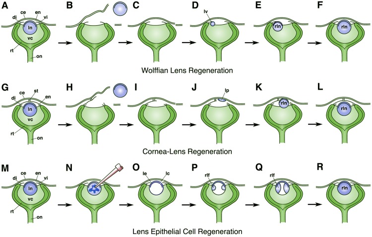
Diagrams illustrating the process of Wolffian lens regeneration (A–F), cornea-lens regeneration (G–L), and lens epithelial cell regeneration (M–R). In (B) and (H), simple lentectomy is performed to remove the intact lens along with its lens capsule. (N) Shows the process of phacoemulsification to remove the lens fiber cells while mainly leaving the lens epithelium and lens capsule intact (as seen in O). (A–F) and (M–R) show adult eyes. Unlike the case in the adult eye, notice that the Xenopus larval cornea epithelium is initially attached to the deeper cornea endothelium by only a small central stalk (as shown in G). This connection enlarges, and the collagenous stroma is deposited during later stages when the larva approaches the time of metamorphosis. Eye structures are labeled as: ce, cornea epithelium; di, dorsal iris; en, cornea endothelium; lc, lens capsule; le, lens epithelium; ln, lens; lp, lens placode; lv, lens vesicle; on, optic nerve; rlf, regenerated lens fiber cells; rln, regenerated lens; rt, retina, st, central stalk; vc, vitreous chamber; vi, ventral iris.
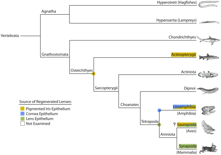
Phylogram showing major vertebrate clades and occurrence of examples that can regenerate the lens. The type of lens regeneration as indicated by different colors, as shown in the key. “?” indicates that reported examples of Wolffian lens regeneration in members of the Sauropsida (i.e., the chicken Gallus gallus) are questionable. Examples from several subphyla or classes, including the more basal groups, have either not yet been examined or reported in the literature. Colored dots represent possible presence of that matching character in the common ancestor for those particular nodes or branches. See text and table 1 for further details. Phylogenetic relationships are based on Meyer and Zardoya (2009).
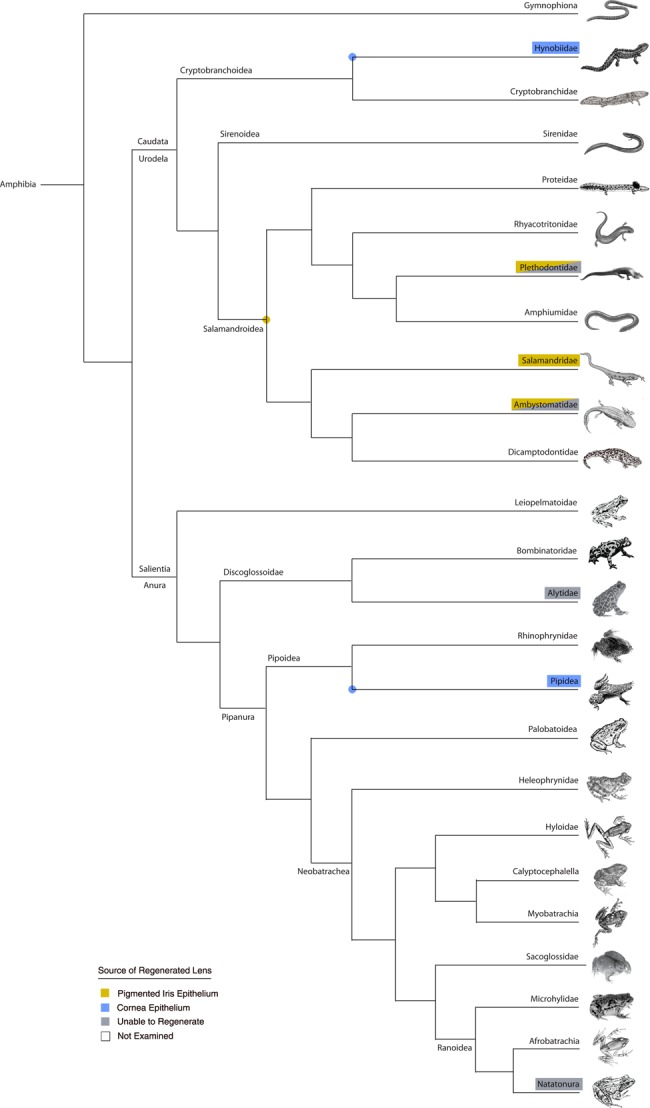
Phylogram showing major amphibian clades and occurrence of verified examples that can and cannot regenerate the lens. The type of lens regeneration is indicated by different colors, as shown in the key. Examples of lens epithelial cell (LEC) regeneration are not mapped onto this particular tree (however, see table 1). Representatives from several families, including the more basal groups, have not yet been examined. Colored dots represent possible presence of that matching character in the common ancestor for those particular nodes or branches. See text and table 1 for further details. Phylogenetic relationships based on Germain and Laurin 2009; Pyron and Wiens 2011; and Feng et al. 2017.
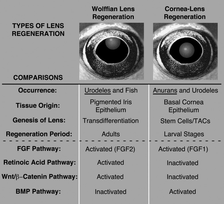
Summary comparing features of Wolffian lens regeneration with cornea-lens regeneration. See text for further explanation. TACs, transit amplifying cells; BMP, bone morphogenetic protein; FGF, fibroblast growth factor; Wnt, Wingless-related integration site.
References
-
- Agarwal LP, Angra SK, Knosla PK, Tandon HD.. 1964. Lens regeneration in mammals: II Monkeys. Orient Arch Opthalmol. 2:47–59.
-
- Ang SJ, Stump RJ, Lovicu FJ, McAvoy JW.. 2004. Spatial and temporal expression of Wnt and Dickkopf genes during murine lens development. Gene Expr Patterns 43:289–295. - PubMed
-
- Arresta E, Bernardini S, Gargioli C, Filoni S, Cannata SM.. 2005. Lens-forming competence in the epidermis of Xenopus laevis during development. J Exp Zool. 303A1:1–12. - PubMed
-
- Barbosa-Sabanero K, Hoffmann A, Judge C, Lightcap N, Tsonis PA, Del Rio-Tsonis K.. 2012. Lens and retina regeneration: new perspectives from model organisms. Biochem J. 4473:321–334. - PubMed
-
- Belecky-Adams TL, Adler R, Beebe DC.. 2002. Bone morphogenetic protein signaling and the initiation of lens fiber cell differentiation. Development 12916:3795–3802. - PubMed
Publication types
MeSH terms
Grants and funding
LinkOut - more resources
Full Text Sources
Other Literature Sources
