Review
. 2018 Jun 7:9:229-252.
doi: 10.1146/annurev-chembioeng-060817-084034. Epub 2018 Mar 26.

Biomolecular Ultrasound and Sonogenetics

Affiliations
Review

Biomolecular Ultrasound and Sonogenetics

David Maresca et al. Annu Rev Chem Biomol Eng. .

Abstract

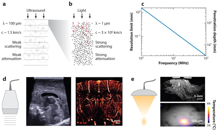
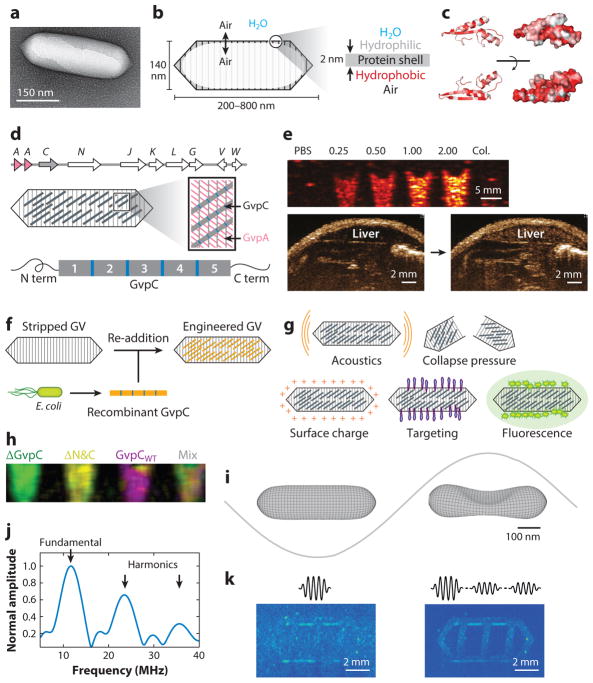
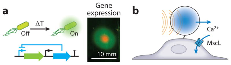
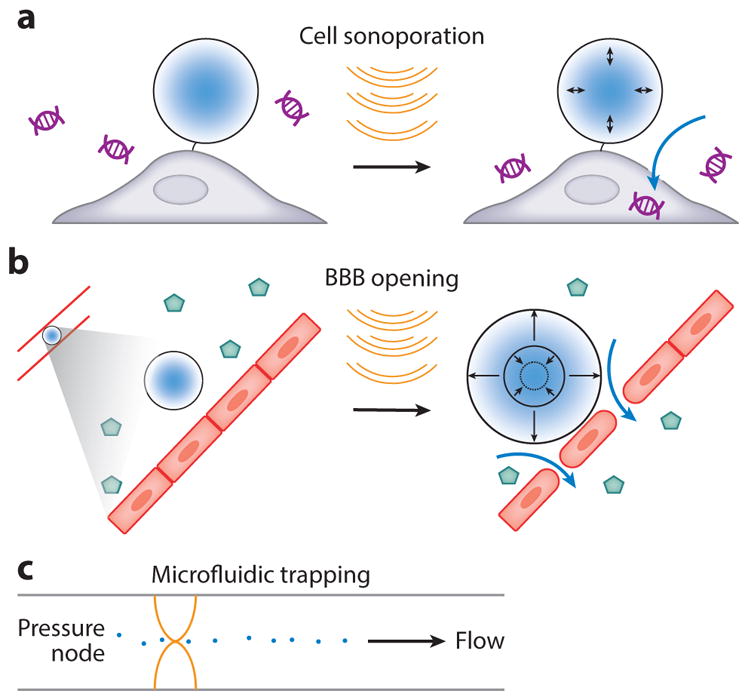
Visualizing and modulating molecular and cellular processes occurring deep within living organisms is fundamental to our study of basic biology and disease. Currently, the most sophisticated tools available to dynamically monitor and control cellular events rely on light-responsive proteins, which are difficult to use outside of optically transparent model systems, cultured cells, or surgically accessed regions owing to strong scattering of light by biological tissue. In contrast, ultrasound is a widely used medical imaging and therapeutic modality that enables the observation and perturbation of internal anatomy and physiology but has historically had limited ability to monitor and control specific cellular processes. Recent advances are beginning to address this limitation through the development of biomolecular tools that allow ultrasound to connect directly to cellular functions such as gene expression. Driven by the discovery and engineering of new contrast agents, reporter genes, and bioswitches, the nascent field of biomolecular ultrasound carries a wave of exciting opportunities.

Keywords: biomolecular; gas vesicles; imaging; reporter gene; sonogenetics; ultrasound.


Figures

Figure 1
Figure 1
Properties and applications of ultrasound waves. (a) Physical properties of ultrasound waves in biological tissues. (b) Physical properties of light traveling in biological tissue. (c) Fundamental tradeoff between ultrasound resolution and penetration depth as a function of frequency in brain tissue (penetration depth was assessed based on a 60-decibel round-trip attenuation). At an ultrasound frequency of 15 MHz, one can expect to image the brain 2 cm deep at a 100-μm resolution. (d) Illustration of ultrasound imaging capabilities; conventional B-mode image of an infant brain with a submillimeter resolution of cerebral structures; 15-MHz superresolution ultrasound image of the rat brain vasculature with an 8-μm resolution, breaking the classical tradeoff exposed in c [adapted with permission from Errico et al. (18)]. (e) Illustration of focused ultrasound energy delivery with millimeter precision. (top) Local blood-brain barrier opening induced by stable microbubble cavitation and tracked with gadolinium-enhanced magnetic resonance image (MRI); (bottom) MRI temperature map of a phantom during focused ultrasound insonification.
Figure 2
Figure 2
Regimes of biomolecular ultrasound. (a) Short ultrasound pulse backscattering is used to image acoustic biomolecules in vivo. (b) Extended ultrasound pulses induce an acoustic radiation force that can actuate ultrasound-sensitive particles. (c) Continuous ultrasound waves lead to local heating and can be used to turn on thermal bioswitches. (d) Low-frequency ultrasound can be used to cavitate microbubbles that can induce cell or vascular barrier disruption.
Figure 3
Figure 3
Gas vesicles as acoustic biomolecules. (a) Transmission electron micrograph of individual gas vesicle (GV) from Anabaena flos-aquae. (b) Illustration of GV structure. (c) Protein folding model for A. flos-aquae gas vesicle protein A (GvpA), colored to indicate hydrophobilicy (red). Structure from Ezzeldin et al. (40), rendered in PyMOL. (d) Gene cluster encoding A. flos-aquae GVs (top), illustration of GvpA and GvpC spatial arrangement (middle), and repeat structure of GvpC protein (bottom). (e) Ultrasound image of GVs at various optical densities and after hydrostatic collapse in vitro. Image of mouse during and after GV administration in vivo, showing contrast in the liver owing to GV accumulation. (f) Illustration of protocol for replacing native GvpC with engineered versions. (g) GV properties that can be modified by substituting engineered versions of GvpC. (h) Multiplexed image of engineered GVs acquired with pressure spectral unmixing. (i) Finite element model of A. flos-aquae GV buckling under acoustic pressure. (j) Predicted acoustic output from buckling GVs. (k) B-mode and amplitude modulated images of gas vesicles arranged in a phantom with linear background scatterers. Panel a adapted with permission from Lu et al. (143). Panels d, fh adapted with permission from Lakshmanan et al. (43). Panel e adapted with permission from Shapiro et al. (35). Panels i–k adapted with permission from Maresca et al. (45).
Figure 4
Figure 4
Acoustic reporter genes. (a) Diagram of engineered ARG1 gene cluster encoding gas vesicle protein A (GvpA) and GvpC from Anabaena flos-aquae and GvpN–U from Bacillus megaterium (top), resulting in ultrasound contrast when expressed in Escherichia coli or Salmonella typhimurium (bottom), compared with bacteria expressing the LuxABCDE operon. (b) Transmission electron microscopy image of E. coli Nissle 1917 expressing ARG1. (c) Illustration of engineered circuit in which ARG expression is driven by a chemical inducer (top), and ultrasound images of E. coli after induction with different amounts of the inducer [isopropyl β-D-1-thiogalactopyranoside (IPTG), bottom]. (d) Normalized ultrasound signal intensity from bacteria induced with different amounts of the inducer, showing the expected circuit output. (e) Anatomical (gray) and background-subtracted contrast (hot colormap) ultrasound image of mouse with colon containing E. coli Nissle 1917 expressing either ARG1 or LuxABCDE in the indicated spatial arrangement. Panels a–e adapted with permission from Bourdeau et al. (46).
Figure 5
Figure 5
Biomolecular applications of focused ultrasound. (a) Schematic of thermal bioswitch controlling gene expression and in vitro patterned gene expression in the region targeted by focused ultrasound. (b) Mechanical actuation of a mechanosensitive channel by exerting acoustic radiation force on a microbubble. Panel a adapted with permission from Piraner & Abedi (71).
Figure 6
Figure 6
Biomolecular transport using ultrasound. (a) Sonoporation of a cell with a bubble, enabling DNA uptake. (b) Microbubble vibration induces loosening of vascular tight junctions, enabling virus uptake through the blood-brain barrier (BBB). (c) Acoustic trapping of particles in a microfluidic channel with standing ultrasound waves.

References

    1. Szabo TL. Diagnostic Ultrasound Imaging: Inside Out. Cambridge, MA: Academic; 2004.
    1. Azhari H. Basics of Biomedical Ultrasound for Engineers. Hoboken, NJ: John Wiley & Sons; 2010.
    1. Cobbold RS. Foundations of Biomedical Ultrasound. Oxford, UK: Oxford Univ. Press; 2006.
    1. Liang H-D, Noble JA, Wells PNT. Recent advances in biomedical ultrasonic imaging techniques. Interface Focus. 2011;1:475–76.
    1. Powers J, Kremkau F. Medical ultrasound systems. Interface Focus. 2011;1:477–89. - PMC - PubMed

Publication types