Review
. 2018 Apr:51:124-131.
doi: 10.1016/j.ceb.2018.03.004. Epub 2018 Mar 23.

The molecular machines that traffic signaling receptors into and out of cilia

Affiliations
Review

The molecular machines that traffic signaling receptors into and out of cilia

Maxence V Nachury. Curr Opin Cell Biol. 2018 Apr.

Abstract

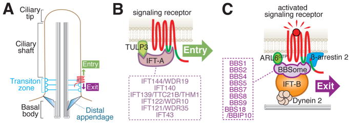
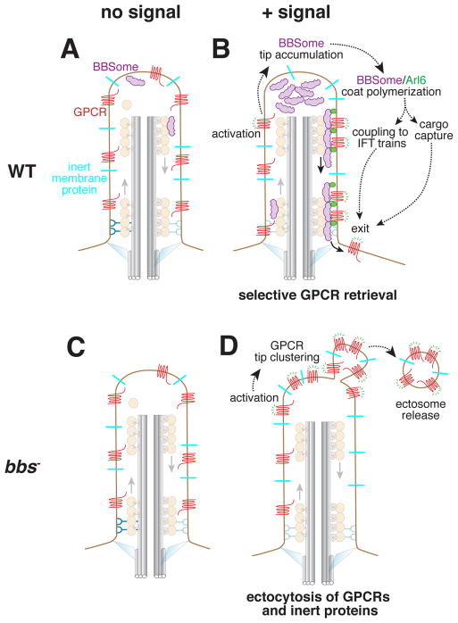
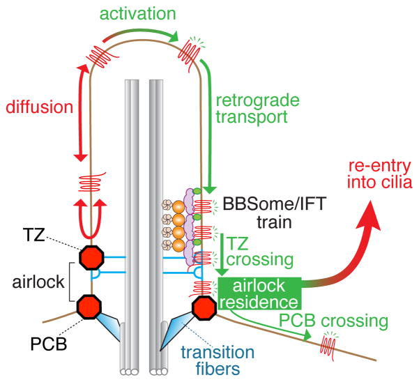
Cilia are surface-exposed organelles that dynamically concentrate signaling molecules to organize sensory, developmental and homeostatic pathways. Entry and exit of signaling receptors is germane to the processing of signals and the molecular machines for entry and exit have started to emerge. The IFT-A complex and its membrane recruitment factor Tulp3 complex promotes the entry of signaling receptors into cilia while the BBSome and its membrane recruitment factor Arl6GTP ferry activated signaling receptors out of cilia. Ciliary exit is a surprisingly complex process entailing passage through a first diffusion barrier at the transition zone, diffusion inside an intermediate compartment and crossing of a periciliary diffusion barrier. The two barriers may organize a privileged compartment where activated signaling receptors transiently reside.


Figures

Figure 1
Figure 1
IFT-A mediates constitutive ciliary entry and the BBSome is in charge of regulated exit from cilia. A. Schematic of relevant ciliary structures. B. Direct binding of the BBSome to the third intracellular loop and the C-terminal tail of various GPCRs has been shown by [10,31,37]. A role for dynein 2 and IFT-B in exit is supported by multiple lines of evidence [17,38]. The IFT-B/Dynein 2 interaction is speculated from the proximity of IFT-B to the microtubule in immuno-electron microscopy [67] and nanoscopy reconstructions [28]. Finally, β-arrestin 2 enables the specific retrieval of activated signaling receptors [34,49,50]. C. Direct binding of IFT-A to the ciliary targeting signal inside the third intracellular loop (green line) of SSTR3 was demonstrated by [34]. Little support is currently available for Kinesin-II and IFT-B participating in the entry of membrane proteins into cilia [45,46] and these complexes were not included in the entry diagram. The subunit composition of IFT-A and BBSome is listed in the boxes.
Figure 2
Figure 2
Conventional and unconventional pathways for exit from cilia. A and C. No exit takes place in the absence of GPCR stimulation. B. Activation of ciliary Gαi-coupled GPCRs (e.g. Sstr3 or Smo) leads to the accumulation of BBSomes at the tip and the Arl6-dependent formation of large and processive retrograde BBSome trains. The requirement for Arl6 in the formation of large trains suggests that these trains are the macroscopic correlates of BBSome/Arl6GTP coats. BBSome-mediated retrieval only removes the activated GPCRs from cilia while leaving ciliary bystanders behind. D. When BBSome or Arl6 function is abrogated, signal-dependent ectocytosis leads to specific removal of activated GPCRs and the non-specific loss of ciliary bystanders [35]. This observation offers a cogent explanation for the loss of several membrane proteins form Bbs mutant cilia.
Figure 3
Figure 3
Two barriers must be crossed for activated GPCRs to exit cilia. The first barrier at the transition zone is crossed by associating with BBSome/IFT trains while the mechanisms for crossing the second barrier remain unknown [34]. Between the transition zone (TZ) and the periciliary barrier (PCB) lies the airlock where GPCR that have crossed the TZ reside for a few seconds before either re-entring cilia (99.5% of the time) or crossing the PCB (0.5% of the time).

References

    1. van Dam TJP, Townsend MJ, Turk M, Schlessinger A, Sali A, Field MC, Huynen MA. Evolution of modular intraflagellar transport from a coatomer-like progenitor. Proc Natl Acad Sci U A. 2013;110:6943–6948. - PMC - PubMed
    1. Cole DG, Diener DR, Himelblau AL, Beech PL, Fuster JC, Rosenbaum JL. Chlamydomonas kinesin-II-dependent intraflagellar transport (IFT): IFT particles contain proteins required for ciliary assembly in Caenorhabditis elegans sensory neurons. J Cell Biol. 1998;141:993–1008. - PMC - PubMed
    1. Nachury MV, Loktev AV, Zhang Q, Westlake CJ, Peränen J, Merdes A, Slusarski DC, Scheller RH, Bazan JF, Sheffield VC, et al. A core complex of BBS proteins cooperates with the GTPase Rab8 to promote ciliary membrane biogenesis. Cell. 2007;129:1201–1213. - PubMed
    1. Loktev AV, Zhang Q, Beck JS, Searby CC, Scheetz TE, Bazan JF, Slusarski DC, Sheffield VC, Jackson PK, Nachury MV. A BBSome subunit links ciliogenesis, microtubule stability, and acetylation. Dev Cell. 2008;15:854–865. - PubMed
    1. Scheidecker S, Etard C, Pierce NW, Geoffroy V, Schaefer E, Muller J, Chennen K, Flori E, Pelletier V, Poch O, et al. Exome sequencing of Bardet-Biedl syndrome patient identifies a null mutation in the BBSome subunit BBIP1 (BBS18) J Med Genet. 2014;51:132–136. - PMC - PubMed

Publication types