. 2018 Mar 27;13(1):42.
doi: 10.1186/s13023-018-0780-z.

Hermansky-Pudlak syndrome type 2 manifests with fibrosing lung disease early in childhood

Affiliations

Hermansky-Pudlak syndrome type 2 manifests with fibrosing lung disease early in childhood

Meike Hengst et al. Orphanet J Rare Dis. .

Abstract

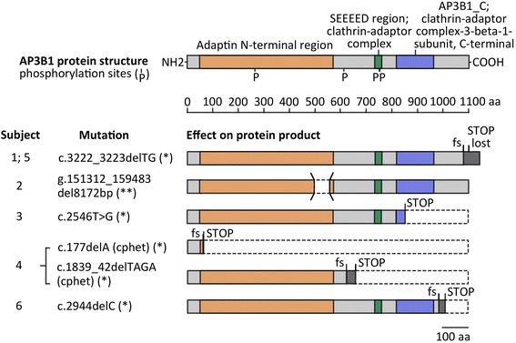
Background: Hermansky-Pudlak syndrome (HPS), a hereditary multisystem disorder with oculocutaneous albinism, may be caused by mutations in one of at least 10 separate genes. The HPS-2 subtype is distinguished by the presence of neutropenia and knowledge of its pulmonary phenotype in children is scarce.

Methods: Six children with genetically proven HPS-2 presented to the chILD-EU register between 2009 and 2017; the data were collected systematically and imaging studies were scored blinded.

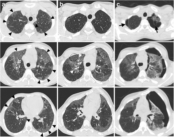
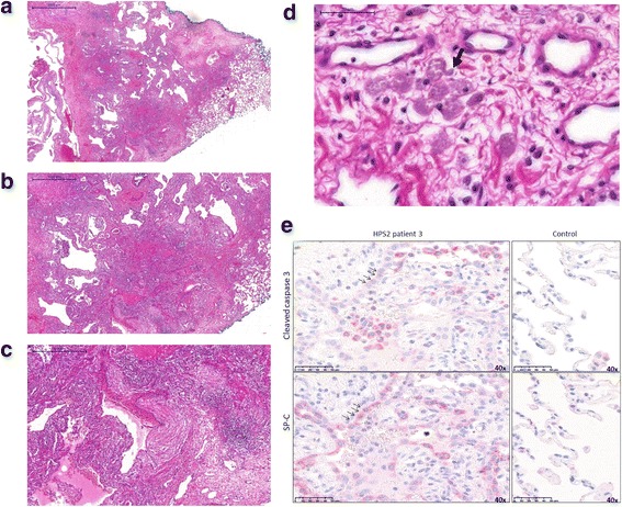
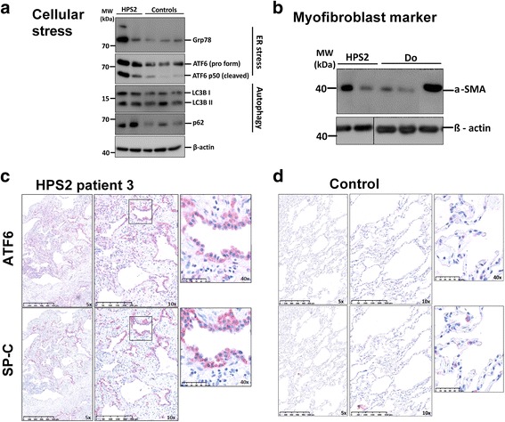
Results: Pulmonary symptoms including dyspnea, coughing, need for oxygen, and clubbing started 3.3 years before the diagnosis was made at the mean age of 8.83 years (range 2-15). All children had recurrent pulmonary infections, 3 had a spontaneous pneumothorax, and 4 developed scoliosis. The frequency of pulmonary complaints increased over time. The leading radiographic pattern was ground-glass opacities with a rapid increase in reticular pattern and traction bronchiectasis between initial and follow-up Computer tomography (CT) in all subjects. Honeycombing and cysts were newly detectable in 3 patients. Half of the patients received a lung biopsy for diagnosis; histological patterns were cellular non-specific interstitial pneumonia, usual interstitial pneumonia-like, and desquamative interstitial pneumonia.

Conclusions: HPS-2 is characterized by a rapidly fibrosing lung disease during early childhood. Effective treatments are required.

Keywords: Childhood; Hermansky-Pudlak syndrome type 2; Pulmonary fibrosis; Pulmonary phenotype; Tachydyspnea.


Conflict of interest statement

Ethics approval and consent to participate

The study was approved by the ethics committee of the Ludwig-Maximilians University of Munich (EK 111-13).

Consent for publication

Informed consent to report individual patient data was obtained by all patients old enough to consent, and their parents or guardians.

Competing interests

The authors declare that they have no competing interests.

Publisher’s Note

Springer Nature remains neutral with regard to jurisdictional claims in published maps and institutional affiliations.

Figures

Fig. 1
Fig. 1
AP3B1 mutations and their predicted effect on protein structure. Homozygous or compound heterozygous mutations found in individuals with HPS-2 are listed and the expected consequence for protein structure is illustrated. Colored segments represent relevant functional protein domains and regions of altered amino acid sequence after reading frame shift (grey). Genetic nomenclature refers to the respective entries in GenBank, NCBI. (*) NM_003664.4 (**) NG_007268.1 Abbreviations: aa = amino acid, cphet = compound heterozygous, fs = frame shift
Fig. 2
Fig. 2
CT images of subject 3. a, b, c CT images at 8.7 years of age (column a) and follow-ups at age of 14.5 and 14.8 (column b and c). Leading pattern is GGO on both sides at initial scan (black arrowheads) and slight increase in reticular markings (encircled) and bronchial wall thickening (white arrowheads) at follow-up. Last follow-up showed distinctive pneumothorax and pleural effusion on the left and subpleural blebs (black arrows) in both lung apices
Fig. 3
Fig. 3
Pulmonary histopathology in subject 3. a-d Histological pattern of patient 3 shows patchy dense interstitial and pleural fibrosis with subpleural cysts/ blebs bordered by normal lung parenchyma at the right margin (c: HE × 10, d: fibrosis and cysts × 20) and small interstitial aggregates of ceroid macrophages with pale brown pigment in the cytoplasm (arrow) as a characteristic histological finding in HPS (D × 400). e Immunohistochemistry identifying alveolar type II cells by staining of proSP-C (SP-C), showing hyperplastic and vacuolated type II pneumocytes as another typical feature of HPS (see also in panel d). Increase in the apoptosis marker cleaved caspase-3 in alveolar epithelial type II cells. Representative images of immunohistochemistry for cleaved caspase-3 and proSP-C performed on serial paraffin sections of HPS-2 and organ donor lungs. Shown here are high magnification images (40×), indicating same type II cells stained for both cleaved caspase-3 and SP-C dying type II cells in HPS-2 patient lungs
Fig. 4
Fig. 4
Activation of ER-stress and autophagy in subject 3. a Western blot images showing increased expression of the endoplasmic reticulum (ER) chaperone GRP78, ER stress marker AFT6, and importantly the p50 (cleaved form) of ATF6 in HPS-2 patient lungs. In addition, the autophagy marker LC3B (active lipidated form is LC3BII) and SQSTM1/p62 are concomitantly increased, indicating defective autophagy in HPS-2 patient lungs (HPS-2 = patient 3, samples from two different sites of a lung, controls = organ donors). b Representative Western blot images for the myofibroblast marker alpha-SMA and loading control, beta-actin in lung homogenates of patient 3 (HPS-2, duplicate lanes with samples from two different sites of a lung), and 3 different organ donors (controls, one lane each). c Increase in ER stress marker ATF6 in alveolar epithelial type II cells in HPS-2 patient lungs. Representative images of immunohistochemistry for ATF6 and proSP-C (SP-C, alveolar epithelial type II cells marker) performed on serial paraffin sections of HPS-2 and organ donor lungs. Shown here are low and high magnification images, using 5×, 10× and 40× objective for both HPS-2 and donor lung sections. Far right images indicate same alveolar epithelial type II cells stained for both ATF6 and SP-C indicating many alveolar epithelial type II cells positively stained for ATF6. Scale bar: as indicated in the images. d Control sections represent ATF6 and SP-C stainings in donor lung sections, where almost no ATF6 is detected in SP-C positive alveolar epithelial type II cells

References

    1. Wildenberg SC, Oetting WS, Almodovar C, Krumwiede M, White JG, King RA. A gene causing Hermansky-Pudlak syndrome in a Puerto Rican population maps to chromosome 10q2. Am J Hum Genet. 1995;57:755–765. - PMC - PubMed
    1. Hurford MT, Sebastiano C. Hermansky-pudlak syndrome: report of a case and review of the literature. Int J Clin Exp Pathol. 2008;1:550–554. - PMC - PubMed
    1. Vicary GW, Vergne Y, Santiago-Cornier A, Young LR, Roman J. Pulmonary fibrosis in Hermansky-Pudlak syndrome. Ann Am Thorac Soc. 2016;13:1839–1846. - PMC - PubMed
    1. Gochuico BR, Huizing M, Golas GA, Scher CD, Tsokos M, Denver SD, Frei-Jones MJ, Gahl WA. Interstitial lung disease and pulmonary fibrosis in Hermansky-Pudlak syndrome type 2, an adaptor protein-3 complex disease. Mol Med. 2012;18:56–64. doi: 10.2119/molmed.2011.00198. - DOI - PMC - PubMed
    1. Araki Y, Ishii Y, Abe Y, Yoshizawa J, Okamoto F, Hozumi Y, Suzuki T. Hermansky-Pudlak syndrome type 4 with a novel mutation. J Dermatol. 2014;41:186–187. doi: 10.1111/1346-8138.12386. - DOI - PubMed

Publication types

Supplementary concepts