. 2018 Apr 4;26(4):1127-1136.
doi: 10.1016/j.ymthe.2018.03.002. Epub 2018 Mar 10.

Dose-Dependent Prevention of Metabolic and Neurologic Disease in Murine MPS II by ZFN-Mediated In Vivo Genome Editing

Affiliations

Dose-Dependent Prevention of Metabolic and Neurologic Disease in Murine MPS II by ZFN-Mediated In Vivo Genome Editing

Kanut Laoharawee et al. Mol Ther. .

Abstract

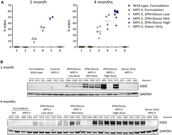
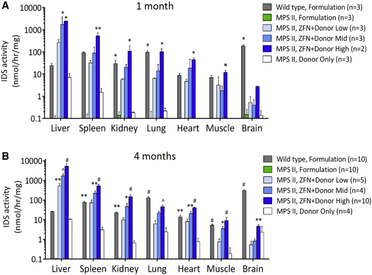
Mucopolysaccharidosis type II (MPS II) is an X-linked recessive lysosomal disorder caused by deficiency of iduronate 2-sulfatase (IDS), leading to accumulation of glycosaminoglycans (GAGs) in tissues of affected individuals, progressive disease, and shortened lifespan. Currently available enzyme replacement therapy (ERT) requires lifelong infusions and does not provide neurologic benefit. We utilized a zinc finger nuclease (ZFN)-targeting system to mediate genome editing for insertion of the human IDS (hIDS) coding sequence into a "safe harbor" site, intron 1 of the albumin locus in hepatocytes of an MPS II mouse model. Three dose levels of recombinant AAV2/8 vectors encoding a pair of ZFNs and a hIDS cDNA donor were administered systemically in MPS II mice. Supraphysiological, vector dose-dependent levels of IDS enzyme were observed in the circulation and peripheral organs of ZFN+donor-treated mice. GAG contents were markedly reduced in tissues from all ZFN+donor-treated groups. Surprisingly, we also demonstrate that ZFN-mediated genome editing prevented the development of neurocognitive deficit in young MPS II mice (6-9 weeks old) treated at high vector dose levels. We conclude that this ZFN-based platform for expression of therapeutic proteins from the albumin locus is a promising approach for treatment of MPS II and other lysosomal diseases.

Keywords: Hunter syndrome; MPS II; albumin locus; gene therapy; iduronate 2-sulfatase; in vivo genome editing; lysosomal disease; zinc finger nuclease.


Figures

Figure 1
Figure 1
Schematic Showing ZFN-Mediated hIDS Gene Transfer and Expression from the Albumin Locus (A) Outline of strategy for ZFN-mediated integration of hIDS at intron 1 of the albumin locus in mouse hepatocytes. (B) Expression cassette of promoterless hIDS cDNA donor construct is shown. A hF9 splice acceptor is followed by the hIDS cDNA sequence and rabbit globin polyadenylation signal flanked on both 5′ and 3′ ends with mouse albumin homology arms and AAV2 ITR sequences.
Figure 2
Figure 2
Albumin Gene Modification and Expression of hIDS in Mouse Hepatocytes (A) Percent indels were determined in DNA extracted from the livers of animals indicated in the key to the right as described in Materials and Methods. Horizontal line represents mean % indels within each group. Dose-dependent ZFN activity was detected in all groups of mice 1 and 4 months after receiving ZFN+donor. Two animals (no. 1264 and no. 1323) showed no ZFN activity, indicating a failure of the injection (see text). Data from animal numbers 1264 and 1323 were thus excluded from all statistical analyses. No ZFN activity was detected in donor only, wild-type, and MPS II control animals. (B) Western blot of hIDS using a human-specific IDS antibody showed consistent levels of hIDS protein correlated to ZFNs % indel activities.
Figure 3
Figure 3
Tissue IDS Activities at 1 Month and 4 Months Post-treatment (A) IDS activities in tissue extracts were determined as described in Materials and Methods. IDS activities increased in a dose-dependent fashion in the ZFN+donor-treated groups at 1 month. (B) At 4 months, IDS activities in all ZFN+donor-treated groups were significantly higher than the MPS II formulation control group and were expressed in a dose-dependent manner. We observed significantly higher IDS activities than wild-type in the liver of all ZFN+donor-treated groups. IDS activities were higher or comparable to wild-type levels in other tissues except brain. IDS activity was not detected in any formulation-treated MPS II tissue. Brain IDS activities in the high-dose group were 1.5% of the wild-type level. Data shown are means ± SEM. *p < 0.05; **p < 0.01; ˆp < 0.001; #p < 0.0001 versus the formulation-treated MPS II control group.
Figure 4
Figure 4
Plasma IDS Activity after Administration of AAV2/8 Delivering ZFNs and hIDS Donor Three different doses of ZFN+donor were injected into each of three different groups of MPS II mice. One group of mice received only hIDS donor (donor only). Wild-type littermates and MPS II littermates were injected with formulation buffer. Plasma was periodically collected post-treatment until day 120 post-injection. Data shown are means ± SEM. *p < 0.05; **p < 0.01; #p < 0.0001 versus wild-type group.
Figure 5
Figure 5
Glycosaminoglycan Content in Tissues Post-injection Levels of tissue GAGs at 1 month (A) and 4 months (B) in MPS II control animals were significantly elevated when compared to wild-type. GAG contents were reduced or normalized in all tissues tested (except brain) in all treated groups at 1 and 4 months post-treatment. *p < 0.05; **p < 0.01; ˆp < 0.001; #p < 0.0001 versus the formulation-treated MPS II control group. (C) Levels of dermatan and heparan sulfate in mouse brain homogenates, 4 months post-injection, are shown. Horizontal lines represent the mean of each group. All wild-type mice had levels of dermatan sulfate at or below the lower limit of quantification for this assay (< 0.005 μg DS/mg protein lysate); *p < 0.05 for high-dose-treated versus untreated MPS II mice. Data shown are means ± SEM. (D) Correction of cellular vacuolation in ZFN+donor-treated MPS II mice is shown. Tissue sections collected from all animals were fixed in 10% neutral-buffered formalin, embedded in paraffin, and processed to slides. Slides were stained with H&E and evaluated by a board-certified veterinary pathologist (Seventh Wave Laboratories) for potential evidence of pathology and degree of tissue vacuolation. Images are shown at 40× magnification. Arrows indicate the location of disease-related microvesiculated mesenchymal cells. A complete list of corrected tissues is in Table S1.
Figure 6
Figure 6
Barnes Maze Performance of High-Dose ZFN+Donor-Treated Group at 4 Months Post-injection Cognitive performance was assessed using the Barnes maze at 4 months post-treatment. (A) Data represent mean ± SEM of the time required to escape the platform over 6 days of testing. Wild-type and high-dose ZFN+donor groups showed significantly better performance when compared to MPS II control animals (p < 0.05; *MPS II formulation versus ZFN+donor high; #MPS II formulation versus wild-type). Performance comparable to wild-type was observed in the high-dose group. Mid-dose, low-dose, and donor-only groups did not perform better than the formulation-treated MPS II control group (data not shown). (B) Data from individual mice on day 6 of testing are shown. Data shown are means ± SEM.

References

    1. Nathwani A.C., Reiss U.M., Tuddenham E.G.D., Rosales C., Chowdary P., McIntosh J., Della Peruta M., Lheriteau E., Patel N., Raj D. Long-term safety and efficacy of factor IX gene therapy in hemophilia B. N. Engl. J. Med. 2014;371:1994–2004. - PMC - PubMed
    1. Bennett J., Ashtari M., Wellman J., Marshall K.A., Cyckowski L.L., Chung D.C., McCague S., Pierce E.A., Chen Y., Bennicelli J.L. AAV2 gene therapy readministration in three adults with congenital blindness. Sci. Transl. Med. 2012;4:120ra15. - PMC - PubMed
    1. Sharma R., Anguela X.M., Doyon Y., Wechsler T., DeKelver R.C., Sproul S., Paschon D.E., Miller J.C., Davidson R.J., Shivak D. In vivo genome editing of the albumin locus as a platform for protein replacement therapy. Blood. 2015;126:1777–1784. - PMC - PubMed
    1. Li H., Haurigot V., Doyon Y., Li T., Wong S.Y., Bhagwat A.S., Malani N., Anguela X.M., Sharma R., Ivanciu L. In vivo genome editing restores haemostasis in a mouse model of haemophilia. Nature. 2011;475:217–221. - PMC - PubMed
    1. Lombardo A., Genovese P., Beausejour C.M., Colleoni S., Lee Y.-L., Kim K.A., Ando D., Urnov F.D., Galli C., Gregory P.D. Gene editing in human stem cells using zinc finger nucleases and integrase-defective lentiviral vector delivery. Nat. Biotechnol. 2007;25:1298–1306. - PubMed

Publication types

MeSH terms