

Deletion of the endogenous TrkB.T1 receptor isoform restores the number of hippocampal CA1 parvalbumin-positive neurons and rescues long-term potentiation in pre-symptomatic mSOD1(G93A) ALS mice
- PMID: 29580900
- PMCID: PMC8108068
- DOI: 10.1016/j.mcn.2018.03.010
Deletion of the endogenous TrkB.T1 receptor isoform restores the number of hippocampal CA1 parvalbumin-positive neurons and rescues long-term potentiation in pre-symptomatic mSOD1(G93A) ALS mice
Abstract
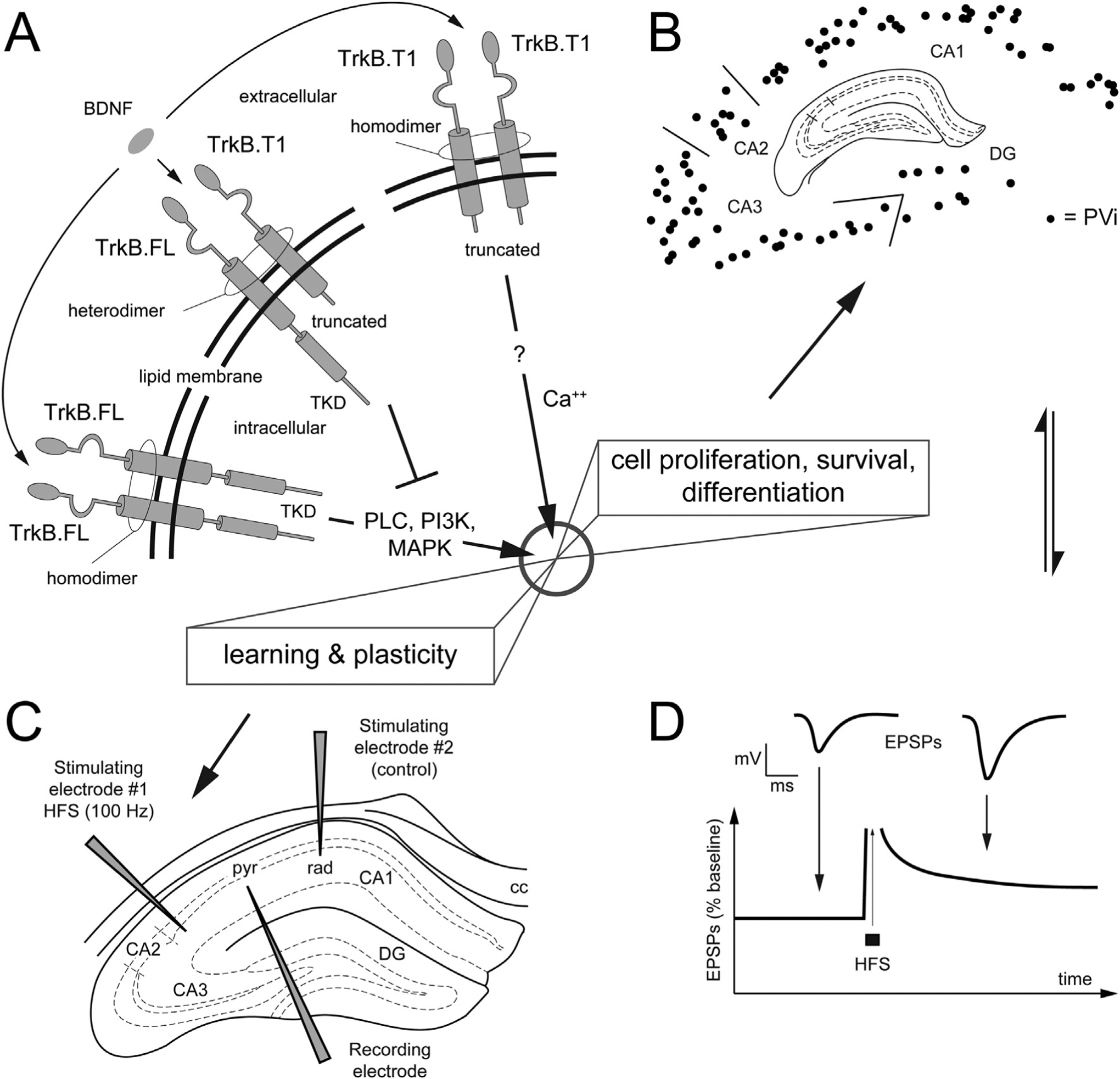
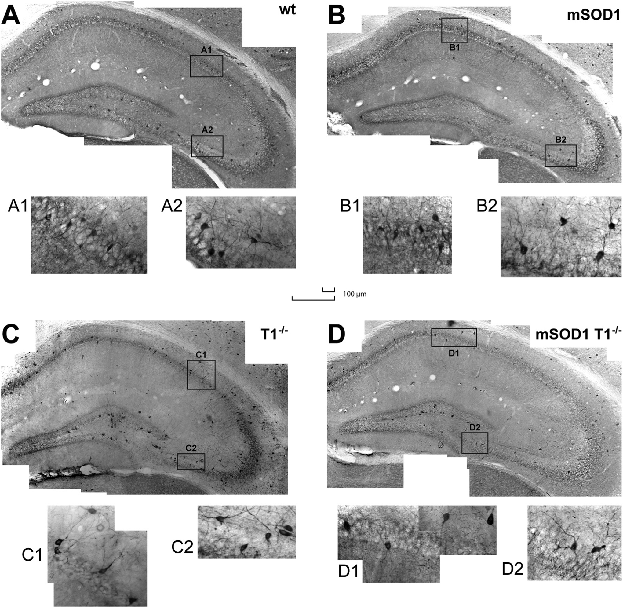
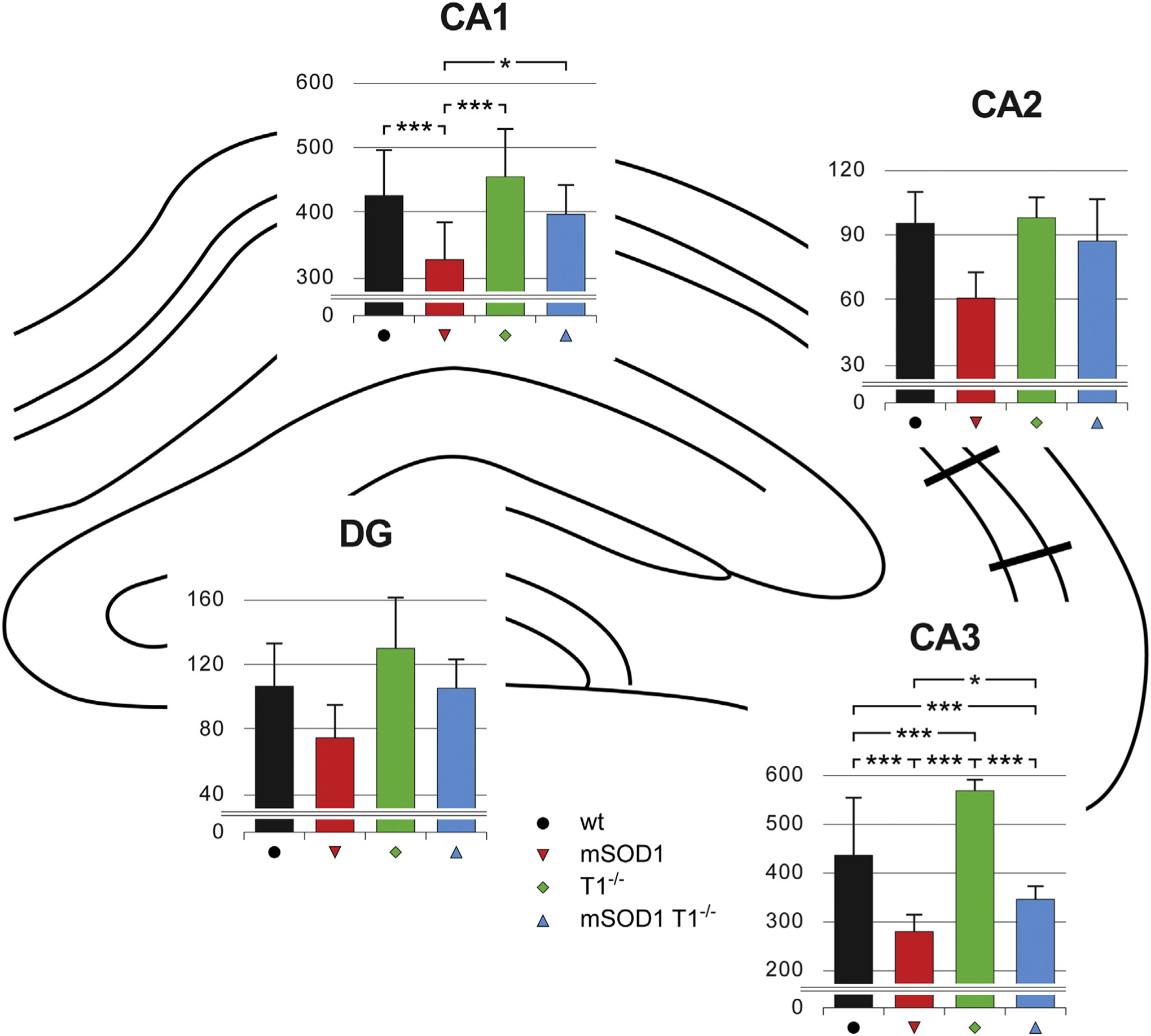
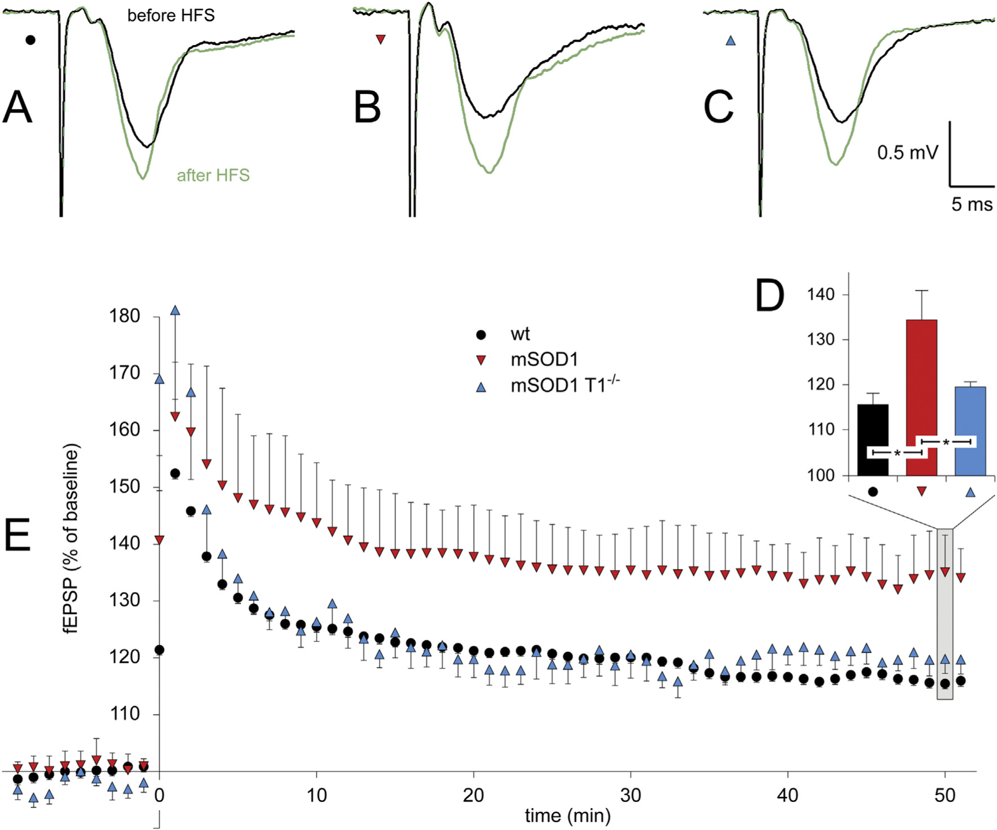
Amyotrophic lateral sclerosis (ALS) causes rapidly progressive paralysis and death within 5 years from diagnosis due to degeneration of the motor circuits. However, a significant population of ALS patients also shows cognitive impairments and progressive hippocampal pathology. Likewise, the mutant SOD1(G93A) mouse model of ALS (mSOD1), in addition to loss of spinal motor neurons, displays altered spatial behavior and hippocampal abnormalities including loss of parvalbumin-positive interneurons (PVi) and enhanced long-term potentiation (LTP). However, the cellular and molecular mechanisms underlying these morpho-functional features are not well understood. Since removal of TrkB.T1, a receptor isoform of the brain-derived neurotrophic factor, can partially rescue the phenotype of the mSOD1 mice, here we tested whether removal of TrkB.T1 can normalize the number of PVi and the LTP in this model. Stereological analysis of hippocampal PVi in control, TrkB.T1-/-, mSOD1, and mSOD1 mice deficient for TrkB.T1 (mSOD1/T1-/-) showed that deletion of TrkB.T1 restored the number of PVi to physiological level in the mSOD1 hippocampus. The rescue of PVi neuron number is paralleled by a normalization of high-frequency stimulation-induced LTP in the pre-symptomatic mSOD1/T1-/- mice. Our experiments identified TrkB.T1 as a cellular player involved in the homeostasis of parvalbumin expressing interneurons and, in the context of murine ALS, show that TrkB.T1 is involved in the mechanism underlying structural and functional hippocampal degeneration. These findings have potential implications for hippocampal degeneration and cognitive impairments reported in ALS patients at early stages of the disease.
Keywords: Amyotrophic lateral sclerosis; Hippocampus; Long-term potentiation; Parvalbumin-positive interneurons; SOD1(G93A) mouse; TrkB.
Copyright © 2018 Elsevier Inc. All rights reserved.
Conflict of interest statement
Disclosures
No conflicts of interest, financial or otherwise, are declared by the authors.
Figures
Similar articles
-
Increased anxiety-like behavior and selective learning impairments are concomitant to loss of hippocampal interneurons in the presymptomatic SOD1(G93A) ALS mouse model.J Comp Neurol. 2015 Aug 1;523(11):1622-38. doi: 10.1002/cne.23759. Epub 2015 Apr 7. J Comp Neurol. 2015. PMID: 25684566
-
Delayed onset of inherited ALS by deletion of the BDNF receptor TrkB.T1 is non-cell autonomous.Exp Neurol. 2021 Mar;337:113576. doi: 10.1016/j.expneurol.2020.113576. Epub 2020 Dec 24. Exp Neurol. 2021. PMID: 33359475 Free PMC article.
-
C-Boutons and Their Influence on Amyotrophic Lateral Sclerosis Disease Progression.J Neurosci. 2021 Sep 22;41(38):8088-8101. doi: 10.1523/JNEUROSCI.0660-21.2021. Epub 2021 Aug 11. J Neurosci. 2021. PMID: 34380764 Free PMC article.
-
Inducible nitric oxide synthase is present in motor neuron mitochondria and Schwann cells and contributes to disease mechanisms in ALS mice.Brain Struct Funct. 2010 Mar;214(2-3):219-34. doi: 10.1007/s00429-009-0226-4. Epub 2009 Nov 4. Brain Struct Funct. 2010. PMID: 19888600 Free PMC article.
-
Transgenic mice with human mutant genes causing Parkinson's disease and amyotrophic lateral sclerosis provide common insight into mechanisms of motor neuron selective vulnerability to degeneration.Rev Neurosci. 2007;18(2):115-36. doi: 10.1515/revneuro.2007.18.2.115. Rev Neurosci. 2007. PMID: 17593875 Review.
Cited by
-
Parvalbumin interneurons dysfunction is potentially associated with FαMNs decrease and NRG1-ErbB4 signaling inhibition in spinal cord in amyotrophic lateral sclerosis.Aging (Albany NY). 2023 Dec 28;15(24):15324-15339. doi: 10.18632/aging.205351. Epub 2023 Dec 28. Aging (Albany NY). 2023. PMID: 38157256 Free PMC article.
-
The Role of Altered BDNF/TrkB Signaling in Amyotrophic Lateral Sclerosis.Front Cell Neurosci. 2019 Aug 13;13:368. doi: 10.3389/fncel.2019.00368. eCollection 2019. Front Cell Neurosci. 2019. PMID: 31456666 Free PMC article. Review.
-
Disinhibition-assisted long-term potentiation in the prefrontal-amygdala pathway via suppression of somatostatin-expressing interneurons.Neurophotonics. 2020 Jan;7(1):015007. doi: 10.1117/1.NPh.7.1.015007. Epub 2020 Feb 14. Neurophotonics. 2020. PMID: 32090134 Free PMC article.
-
Function and Mechanisms of Truncated BDNF Receptor TrkB.T1 in Neuropathic Pain.Cells. 2020 May 11;9(5):1194. doi: 10.3390/cells9051194. Cells. 2020. PMID: 32403409 Free PMC article. Review.
-
Effects of Exercise on Long-Term Potentiation in Neuropsychiatric Disorders.Adv Exp Med Biol. 2020;1228:439-451. doi: 10.1007/978-981-15-1792-1_30. Adv Exp Med Biol. 2020. PMID: 32342476 Review.
References
-
- Abdulla S, Machts J, Kaufmann J, Patrick K, Kollewe K, Dengler R, Heinze HJ, Petri S, Vielhaber S, Nestor PJ, 2014. Hippocampal degeneration in patients with amyotrophic lateral sclerosis. Neurobiol. Aging 35, 2639–2645. - PubMed
-
- Abrahams S, Leigh PN, Goldstein LH, 2005. Cognitive change in ALS: a prospective study. Neurology 64, 1222–1226. - PubMed
-
- Angelov B, Angelova A, 2017. Nanoscale clustering of the neurotrophin receptor TrkB revealed by super-resolution STED microscopy. Nano 9, 9797–9804. - PubMed
-
- Angelova A, Angelov B, Drechsler M, Lesieur S, 2013. Neurotrophin delivery using nanotechnology. Drug Discov. Today 18, 1263–1271. - PubMed
Publication types
MeSH terms
Substances
Grants and funding
LinkOut - more resources
Full Text Sources
Other Literature Sources
Medical
Molecular Biology Databases
Miscellaneous
