Case Reports
. 2018 Jun;1864(6 Pt A):2108-2118.
doi: 10.1016/j.bbadis.2018.03.020. Epub 2018 Mar 24.

19q13.12 microdeletion syndrome fibroblasts display abnormal storage of cholesterol and sphingolipids in the endo-lysosomal system

Affiliations
Case Reports

19q13.12 microdeletion syndrome fibroblasts display abnormal storage of cholesterol and sphingolipids in the endo-lysosomal system

Kexin Zhao et al. Biochim Biophys Acta Mol Basis Dis. 2018 Jun.

Abstract

Microdeletions in 19q12q13.12 cause a rare and complex haploinsufficiency syndrome characterized by intellectual deficiency, developmental delays, and neurological movement disorders. Variability in the size and interval of the deletions makes it difficult to attribute the complex clinical phenotype of this syndrome to an underlying gene(s). As an alternate approach, we examined the biochemical and metabolic features of fibroblasts from an affected individual to derive clues as to the molecular basis for the syndrome. Immunofluorescence and electron microscopy of affected fibroblasts revealed an abnormal endo-lysosomal compartment that was characterized by rapid accumulation of lysosomotropic dyes, elevated LAMP1 and LAMP2 expression and vacuoles containing membrane whorls, common features of lysosomal lipid storage disorders. The late endosomes-lysosomes (LE/LY) of affected fibroblasts accumulated low-density lipoprotein cholesterol, and displayed reduced cholesterol esterification and increased de novo cholesterol synthesis, indicative of defective cholesterol transport to the endoplasmic reticulum. Affected fibroblasts also had increased ceramide and sphingolipid mass, altered glycosphingolipid species and accumulation of a fluorescent lactosylceramide probe in LE/LY. Autophagosomes also accumulated in affected fibroblasts because of decreased fusion with autolysosomes, a defect associated with other lysosomal storage diseases. Attempts to correct the cholesterol/sphingolipid storage defect in fibroblasts with cyclodextrin, sphingolipid synthesis inhibitors or by altering ion transport were unsuccessful. Our data show that 19q13.12 deletion fibroblasts have abnormal accumulation of cholesterol and sphingolipids in the endo-lysosomal system that compromises organelle function and could be an underlying cause of the clinical features of the syndrome.

Keywords: Autophagy; Cholesterol; Chromosome 19 deletion syndrome; Lipid storage disorder; Lysosomes; Sphingolipids.


Figures

Figure 1.
Figure 1.
Altered expression and processing of LE/LY proteins in 19MD fibroblasts. (A) Control and 19MD fibroblasts were immunostained for LAMP1 and Rab7 using AlexaFluor-488 and −594 secondary antibodies, respectively. Wide field images were captured described in Materials and Methods. (B, C and D) Whole cell lysates from control (C1–C5), Niemann-Pick A (NPA), Niemann-Pick C (NPC) and 19MD fibroblasts were immunoblotted for LAMP1 and LAMP2 and quantified relative to actin (results are from five experiments that included all eight cell lines) (E) Immunoblot analysis of NPC1, ACAT1 and LAMP1 in control and 19MD fibroblasts. (F and G) Immunoblot analysis of mature (M) and processed (P) forms of cathepsin D in control and 19MD fibroblasts quantified relative to actin (results are from three experiments that included all three cell lines). Results in panels C, D and G are the mean and SD of the indicated number of experiments (multiple comparisons, one-way ANOVA, *p<0.05, **p<0.01, ***p<0.001, ****p<0.0001; ns, not significant.).
Figure 2.
Figure 2.
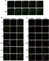
Comparative imaging of lysosomes in 19MD and Niemann-Pick fibroblasts. (A) Time course of LysoTracker DND-26 uptake (20 nM) by live fibroblasts. (B) Control and affected fibroblasts were incubated with pHrodo-Red Dextran (10 mg/ml, 45 min) and LysoTracker green DND-26 (20 nM, 30 min) Images of live cells were captured by spinning disc confocal microscopy as described in the Materials and Methods. (C) Control and affected fibroblasts were cultured in DQ-red-BSA (10 μM). After 6 h, cells were fixed and immunostained with LAMP2 and Alexa Fluor-488 antibodies. Images are confocal sections (0.7 μm, bar 20 μm). The results in each panel are from a representative experiment that was repeated two other times with similar results.
Figure 3.
Figure 3.
Lipid accumulation in the LE/LY of 19MD fibroblasts. (A and B) TEM images of control fibroblasts (C2) cultured in medium A (bar, 1μm in panel A and 200 nm in panel B). (C and D) Low magnification images of 19MD fibroblasts (bar 1 μm). (E) An enlargement of the box area shown in panel D (bar, 200 nm). (F) High magnification image of an 19MD cell showing membranous whorls in the interior of an endosomal vesicle (bar, 200 nm). Results are representative of two experiments.
Figure 4.
Figure 4.
Defective LDL-cholesterol esterification and cholesterol retention in the LE/LY of 19MD fibroblasts. (A) Control and 19MD fibroblasts were cultured in FCS (+LDL) or delipidated serum (−LDL) for 16 h. Cells were fixed, stained with filipin and imaged using a 40x objective. (B and C) Control (C1–C2) and 19MD fibroblasts were cultured as described in panel A followed by treated with 25-hydroxycholesterol (25OH, 2.5 μg/ml) or control solvent for 4 h. [3H]Oleate incorporation into cholesterol ester (CE, panel B) and triacylglycerol (TAG, panel C) was quantified as described in Materials and Methods. Results are the mean and SD of six experiments involving all three cell lines. (D and E) [3H]Oleate incorporation into CE and TAG was measured in control (C3–C5), Niemann-Pick A (NPA), Niemann-Pick C (NPC) and 19MD fibroblasts cultured in DMEM with 10% FCS. Results are the mean and SD of three experiments that included all five cell lines. Statistical comparison in panels B, D and E used multiple comparison, one-way ANOVA (*p<0.01, **p<0.001, ***p< 0.0001; ns, not significant).
Figure 5.
Figure 5.
Elevated sterol synthesis in 19MD fibroblasts. (A) Control (C2) and 19MD fibroblasts were cultured in DMEM with 10% FCS (+LDL) or 10% LPDS (−LDL) for 16 h. Cells were then pulse-labeled with [3H]acetate (10 μCi/ml), and isotope incorporation into cholesterol, lanosterol and fatty acids was quantified as described in Materials and Methods. Results are the mean and SD of five experiments (two-tailed t-test, *p<0.05, **p<0.01; compared to similarly treated control fibroblasts). (B) Cholesterol mass was measured in four control and 19MD fibroblasts (cultured in medium with 10% FCS) by LC-MS/MS as described in Materials and Methods. Results are the mean and SE of three experiments that included all five cell lines (two-way ANOVA, ***p<0.0001 compared to controls).
Figure 6.
Figure 6.
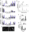
Sphingolipid accumulation in the LE/LY of 19MD fibroblasts. (A) Sphingolipids in control and 19MD fibroblasts (cultured in medium A) were extracted and quantified by LC-MS/MS as described in Materials and Methods. Results are the mean and SE of triplicate measurements in two independent experiments that included all five cell lines (two-tailed t-test; *p<0.05 compared to all controls). (B and C) HPLC elution profiles and relative quantification of anthranilic acid-labeled glycosphingolipid headgroups prepared from control (C2) and 19MD fibroblasts. Results are the mean and SD of three experiments (*p<0.01, **p<0.0001). (D) Incorporation of [3H]serine (10 μCi/ml for 16 h) into sphingolipids in control (C1) and 19MD fibroblasts cultured in medium A. Results are the mean and SD of six experiments (two-tailed t-test; *p<0.05, **p<0.001). (E) Control (C1) and 19MD fibroblasts were incubated in DMEM containing BODIPY-LacCer (2.5 μM), fixed and imaged using a 63x objective as described in Materials and Methods.
Figure 7.
Figure 7.
Defective autophagy in 19MD fibroblasts. (A) Fibroblasts were cultured in medium A or Hank’s buffered salt solution (HBSS) in the presence or absence of chloroquine (50 μM) for 12 h. Total cell lysates were prepared and immunoblotted for LC3 and actin (expression of LC3-II was normalized to actin). Results are the mean and SD of three experiments. B, Autophagosomes (yellow puncta) and autolysosomes (red puncta) were quantified in control and 19MD fibroblasts transduced with lentivirus encoding mCherry-GFP-LC3. Results are shown for 60–70 cells (three experiments) as box and whisker plots (boxes denote the interquartile range with bars at the median and whiskers indicating the 5 and 95 percentiles; *p<0.01 by 2-way ANOVA).
Figure 8.
Figure 8.
Cholesterol and sphingolipid depletion strategies did not correct the storage defect in 19MD fibroblasts. (A and B) 19MD fibroblasts cells were cultured in medium A for 24 h prior to exchange into medium with 10% LPDS and the indicted concentrations of HPBCD for 18 h. Cholesterol and cholesterol esters were extracted and quantified by LC-MS/MS as described in Materials and Methods. Results are the mean and SD of three measurements (two-tailed t-test: *p>0.05, **p<0.01 compared to untreated cells). (C) 19MD fibroblasts received no addition (NA) or 0.3 mM HPBCD for 18 h. Sphingolipids were extracted and quantified by LC-MS/MS. Results are the mean and SD of three measurements (two-tailed t-test: *p>0.05, **p<0.01 compared to untreated cells). (D and E) 19MD and C2 control fibroblasts were cultured in medium A with solvent (no addition, NA), myrocin (myr, 10 μM) or fumonsin B1 (FB, 5 μM) for 16 h. CE and TAG synthesis was measured by pulsing-labeling cells with [3H]oleate. Results are the mean a SD of three experiments. (F and G) 19MD and C2 fibroblasts were incubated in medium A with the indicated concentrations of chloroquine for 48 h prior to measurement of CE and TAG synthesis. Results are the mean and SD of triplicate determinations from a representative experiment.

Similar articles

Cited by

  • Histone lysine methyltransferase-related neurodevelopmental disorders: current knowledge and saRNA future therapies.
    Roth C, Kilpinen H, Kurian MA, Barral S. Roth C, et al. Front Cell Dev Biol. 2023 Feb 27;11:1090046. doi: 10.3389/fcell.2023.1090046. eCollection 2023. Front Cell Dev Biol. 2023. PMID: 36923252 Free PMC article. Review.
  • KMT2B-related disorders: expansion of the phenotypic spectrum and long-term efficacy of deep brain stimulation.
    Cif L, Demailly D, Lin JP, Barwick KE, Sa M, Abela L, Malhotra S, Chong WK, Steel D, Sanchis-Juan A, Ngoh A, Trump N, Meyer E, Vasques X, Rankin J, Allain MW, Applegate CD, Isfahani SA, Baleine J, Balint B, Bassetti JA, Baple EL, Bhatia KP, Blanchet C, Burglen L, Cambonie G, Seng EC, Bastaraud SC, Cyprien F, Coubes C, d'Hardemare V; Deciphering Developmental Disorders Study; Doja A, Dorison N, Doummar D, Dy-Hollins ME, Farrelly E, Fitzpatrick DR, Fearon C, Fieg EL, Fogel BL, Forman EB, Fox RG; Genomics England Research Consortium; Gahl WA, Galosi S, Gonzalez V, Graves TD, Gregory A, Hallett M, Hasegawa H, Hayflick SJ, Hamosh A, Hully M, Jansen S, Jeong SY, Krier JB, Krystal S, Kumar KR, Laurencin C, Lee H, Lesca G, François LL, Lynch T, Mahant N, Martinez-Agosto JA, Milesi C, Mills KA, Mondain M, Morales-Briceno H; NIHR BioResource; Ostergaard JR, Pal S, Pallais JC, Pavillard F, Perrigault PF, Petersen AK, Polo G, Poulen G, Rinne T, Roujeau T, Rogers C, Roubertie A, Sahagian M, Schaefer E, Selim L, Selway R, Sharma N, Signer R, Soldatos AG, Stevenson DA, Stewart F, Tchan M; Undiagnosed Diseases Network; Verma IC, de Vries BBA, Wilson JL, Wong DA, Zaitoun R, Zhen D, Znaczko A, Dale… See abstract for full author list ➔ Cif L, et al. ArXiv [Preprint]. 2025 Feb 10:arXiv:2502.06320v1. ArXiv. 2025. Update in: Brain. 2020 Dec 5;143(11):3242-3261. doi: 10.1093/brain/awaa304. PMID: 39990802 Free PMC article. Updated. Preprint.
  • Construction of copy number variation landscape and characterization of associated genes in a Bangladeshi cohort of neurodevelopmental disorders.
    Akter H, Rahman MM, Sarker S, Basiruzzaman M, Islam MM, Rahaman MA, Rahaman MA, Eshaque TB, Dity NJ, Sarker S, Amin MR, Hossain MM, Lopa M, Jahan N, Hossain S, Islam A, Mondol A, Faruk MO, Saha N, Kundu GK, Kanta SI, Kazal RK, Fatema K, Rahman MA, Hasan M, Hossain Mollah MA, Hosen MI, Karuvantevida N, Begum G, Zehra B, Nassir N, Nabi AHMN, Uddin KMF, Uddin M. Akter H, et al. Front Genet. 2023 Mar 7;14:955631. doi: 10.3389/fgene.2023.955631. eCollection 2023. Front Genet. 2023. PMID: 36959829 Free PMC article.
  • KMT2B-related disorders: expansion of the phenotypic spectrum and long-term efficacy of deep brain stimulation.
    Cif L, Demailly D, Lin JP, Barwick KE, Sa M, Abela L, Malhotra S, Chong WK, Steel D, Sanchis-Juan A, Ngoh A, Trump N, Meyer E, Vasques X, Rankin J, Allain MW, Applegate CD, Attaripour Isfahani S, Baleine J, Balint B, Bassetti JA, Baple EL, Bhatia KP, Blanchet C, Burglen L, Cambonie G, Seng EC, Bastaraud SC, Cyprien F, Coubes C, d'Hardemare V; Deciphering Developmental Disorders Study; Doja A, Dorison N, Doummar D, Dy-Hollins ME, Farrelly E, Fitzpatrick DR, Fearon C, Fieg EL, Fogel BL, Forman EB, Fox RG; Genomics England Research Consortium; Gahl WA, Galosi S, Gonzalez V, Graves TD, Gregory A, Hallett M, Hasegawa H, Hayflick SJ, Hamosh A, Hully M, Jansen S, Jeong SY, Krier JB, Krystal S, Kumar KR, Laurencin C, Lee H, Lesca G, François LL, Lynch T, Mahant N, Martinez-Agosto JA, Milesi C, Mills KA, Mondain M, Morales-Briceno H; NIHR BioResource; Ostergaard JR, Pal S, Pallais JC, Pavillard F, Perrigault PF, Petersen AK, Polo G, Poulen G, Rinne T, Roujeau T, Rogers C, Roubertie A, Sahagian M, Schaefer E, Selim L, Selway R, Sharma N, Signer R, Soldatos AG, Stevenson DA, Stewart F, Tchan M; Undiagnosed Diseases Network; Verma IC, de Vries BBA, Wilson JL, Wong DA, Zaitoun R, Zhen D, Znacz… See abstract for full author list ➔ Cif L, et al. Brain. 2020 Dec 5;143(11):3242-3261. doi: 10.1093/brain/awaa304. Brain. 2020. PMID: 33150406 Free PMC article.
  • Update on KMT2B-Related Dystonia.
    Zech M, Lam DD, Winkelmann J. Zech M, et al. Curr Neurol Neurosci Rep. 2019 Nov 25;19(11):92. doi: 10.1007/s11910-019-1007-y. Curr Neurol Neurosci Rep. 2019. PMID: 31768667 Review.

References

    1. Chowdhury S, Bandholz AM, Parkash S, Dyack S, Rideout AL, Leppig KA, Thiese H, Wheeler PG, Tsang M, Ballif BC, Shaffer LG, Torchia BS, Ellison JW, Rosenfeld JA, Phenotypic and molecular characterization of 19q12q13.1 deletions: a report of five patients, Am. J. Med. Genet. A 164A (2014) 62–69. - PubMed
    1. Malan V, Raoul O, Firth HV, Royer G, Turleau C, Bernheim A, Willatt L, Munnich A, Vekemans M, Lyonnet S, Cormier-Daire V, Colleaux L, 19q13.11 deletion syndrome: a novel clinically recognisable genetic condition identified by array comparative genomic hybridisation, J. Med. Genet 46 (2009) 635–640. - PubMed
    1. Gana S, Veggiotti P, Sciacca G, Fedeli C, Bersano A, Micieli G, Maghnie M, Ciccone R, Rossi E, Plunkett K, Bi W, Sutton VR, Zuffardi O, 19q13.11 cryptic deletion: description of two new cases and indication for a role of WTIP haploinsufficiency in hypospadias, Eur. J. Hum. Genet 20 (2012) 852–856. - PMC - PubMed
    1. Meyer E, Carss KJ, Rankin J, Nichols JM, Grozeva D, Joseph AP, Mencacci NE, Papandreou A, Ng J, Barral S, Ngoh A, Ben-Pazi H, Willemsen MA, Arkadir D, Barnicoat A, Bergman H, Bhate S, Boys A, Darin N, Foulds N, Gutowski N, Hills A, Houlden H, Hurst JA, Israel Z, Kaminska M, Limousin P, Lumsden D, McKee S, Misra S, Mohammed SS, Nakou V, Nicolai J, Nilsson M, Pall H, Peall KJ, Peters GB, Prabhakar P, Reuter MS, Rump P, Segel R, Sinnema M, Smith M, Turnpenny P, White SM, Wieczorek D, Wiethoff S, Wilson BT, Winter G, Wragg C, Pope S, Heales SJ, Morrogh D, Consortium UK, Deciphering Developmental Disorders S, Consortium NBRD, Pittman A, Carr LJ, Perez-Duenas B, Lin JP, Reis A, Gahl WA, Toro C, Bhatia KP, Wood NW, Kamsteeg EJ, Chong WK, Gissen P, Topf M, Dale RC, Chubb JR, Raymond FL, Kurian MA, Mutations in the histone methyltransferase gene KMT2B cause complex early-onset dystonia, Nat. Genet 49 (2017) 223–237. - PubMed
    1. Schuurs-Hoeijmakers JH, Vermeer S, van Bon BW, Pfundt R, Marcelis C, de Brouwer AP, de Leeuw N, de Vries BB, Refining the critical region of the novel 19q13.11 microdeletion syndrome to 750 Kb, J. Med. Genet 46 (2009) 421–423. - PubMed

Publication types

MeSH terms