. 2018 Jun 1;293(22):8543-8553.
doi: 10.1074/jbc.M117.789305. Epub 2018 Mar 26.

Intestinal mucin activates human dendritic cells and IL-8 production in a glycan-specific manner

Affiliations

Intestinal mucin activates human dendritic cells and IL-8 production in a glycan-specific manner

Felipe Melo-Gonzalez et al. J Biol Chem. .

Abstract

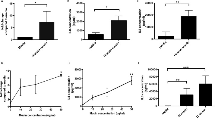
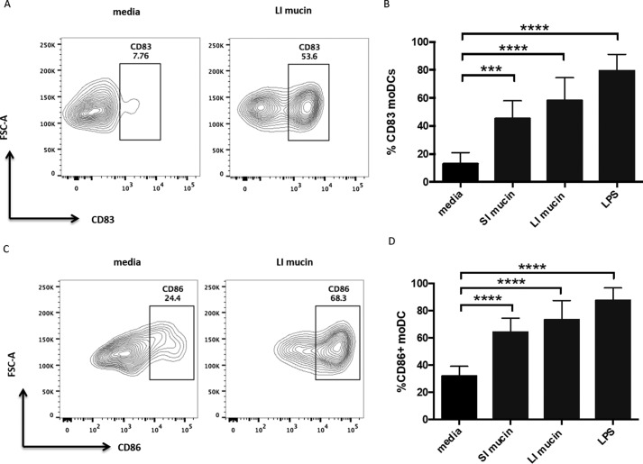
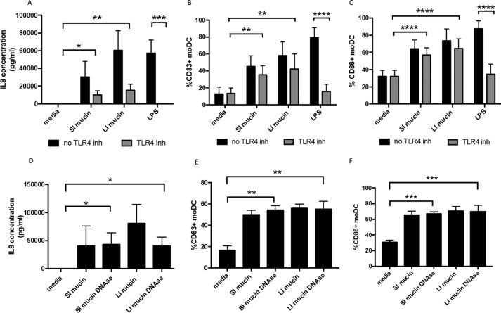
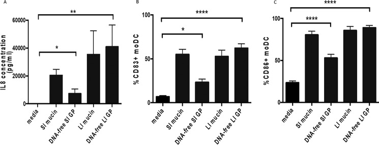
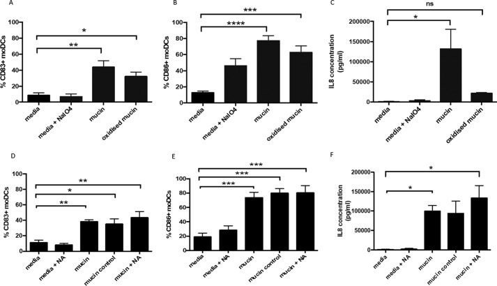
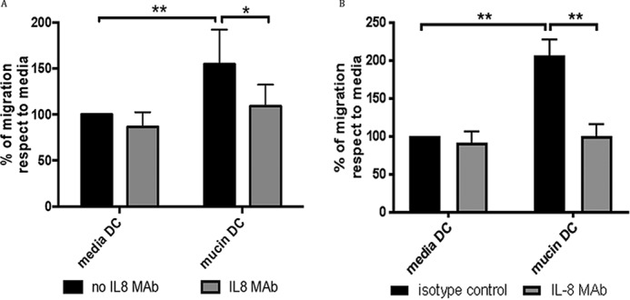
Cross-talk between different components of the intestinal barrier and the immune system may be important in maintaining gut homeostasis. A crucial part of the gut barrier is the mucus layer, a cross-linked gel on top of the intestinal epithelium that consists predominantly of the mucin glycoprotein MUC2. However, whether the mucin layer actively regulates intestinal immune cell responses is not clear. Because recent evidence suggests that intestinal dendritic cells (DCs) may be regulated by the mucus layer, we purified intestinal mucin, incubated it with human DCs, and determined the functional effects. Here we show that expression of the chemokine IL-8 and co-stimulatory DC markers CD86 and CD83 are significantly up-regulated on human DCs in the presence of intestinal mucins. Additionally, mucin-exposed DCs promoted neutrophil migration in an IL-8-dependent manner. The stimulatory effects of mucins on DCs were not due to mucin sample contaminants such as lipopolysaccharide, DNA, or contaminant proteins. Instead, mucin glycans are important for the pro-inflammatory effects on DCs. Thus, intestinal mucins are capable of inducing important pro-inflammatory functions in DCs, which could be important in driving inflammatory responses upon intestinal barrier damage.

Keywords: dendritic cell; inflammation; intestine; mucin; mucosal immunology.


Conflict of interest statement

The authors declare that they have no conflicts of interest with the contents of this article

Figures

Figure 1.
Figure 1.
Human and mouse intestinal mucin can promote IL-8 production on moDCs. Human moDCs were treated overnight with human secreted mucin (2 or 50 μg/ml) and different concentrations of mouse large intestinal mucin (10, 25, and 50 μg/ml). IL-8 expression was measured by qPCR and ELISA. A and B, moDCs were stimulated overnight with human secreted mucin (2 μg/ml) and IL-8 expression measured by qPCR (A) and ELISA (B). n ≥ 5 independent experiments; *, p < 0.05 assessed using unpaired Student's t test. C, ELISA was performed to detect IL-8 in supernatants from untreated and human secreted mucin (50 μg/ml)-treated moDCs. n = 6 independent experiments; **, p < 0.01 assessed using unpaired Student's t test. D and E, moDCs were treated with increasing concentrations of mouse large intestinal mucin (10, 25, and 50 μg/ml) and IL-8 measured by qPCR (D, n = 3; *, p < 0.05 assessed using one-way ANOVA with Dunnet's multiple comparison test) and ELISA (E, n = 5; **, p < 0.01 assessed via Kruskal–Wallis test with Dunn's multiple comparison test). F, moDCs were stimulated overnight with mouse small intestinal (SI) and large intestinal (LI) mucins (50 μg/ml both). ELISA was performed to detect IL-8 on supernatants from untreated and mucin-treated moDCs. n = 8. **, p < 0.01; ***, p < 0.001 assessed using Kruskal–Wallis test with Dunn's multiple comparison test.
Figure 2.
Figure 2.
Mouse intestinal mucin can induce up-regulation of DC activation markers. Human moDC were treated with mucin (50 μg/ml) or LPS (10 ng/ml), and activation markers were detected by flow cytometry. A and C, representative flow cytometry plots showing moDC expression of CD83 (A) and CD86 (C) either untreated or treated with large intestinal (LI) mucin. B and D, percentages of CD83+ moDCs (B) and CD86+ moDCs (D) untreated and treated with small intestinal mucin (SI), large intestinal mucin (LI), and LPS. n = 8 independent experiments. ***, p < 0.001; ****, p < 0.0001 assessed via one-way ANOVA followed by Dunnet's multiple comparison test.
Figure 3.
Figure 3.
Mucin-induced DC activation and IL-8 production are independent of LPS and DNA. moDCs were treated with small intestinal (SI) and large intestinal (LI) mucins (50 μg/ml) in the absence (black bars) and the presence (gray bars) of TLR4 inhibitor CLI-095. LPS (10 ng/ml) was used as a control. A, ELISA was performed to detect IL-8 in supernatants from untreated and mucin-treated moDCs. n = 8 independent experiments. *, p < 0.05; **, p < 0.01; ***, p < 0.001 assessed using Kruskal–Wallis test followed by Dunn's multiple comparison test. B and C, percentages of CD83+ (B) and CD86+ (C) moDCs either untreated or treated with mucin (SI and LI) or LPS, in the presence of the TLR4 inhibitor. n = 8 independent experiments. **, p < 0.01; ****, p < 0.0001 assessed using one-way ANOVA followed by Dunnet's multiple comparison test. D, detection of IL-8 in supernatants from moDCs either left untreated or treated with mucin or DNase-treated mucin (SI and LI, 50 μg/ml) by ELISA. n = 4 independent experiments. *, p < 0.05 assessed using Kruskal–Wallis test followed by Dunn's multiple comparison test. E and F, percentages of CD83+ (E) and CD86+ (F) moDCs either left untreated or treated with mucin or DNase-treated mucin (SI and LI). n = 4 independent experiments. **, p < 0.01; ***, p < 0.001 assessed using one-way ANOVA followed by Dunnet's multiple comparison test.
Figure 4.
Figure 4.
DNA-free mucin glycopeptides induce IL-8 production and DC activation. moDCs were treated with small intestinal (SI) and large intestinal (LI) mucins (50 μg/ml) and mucin glycopeptides (50 μg/ml, previously treated with DNase). A, detection of IL-8 in supernatants from moDCs either left untreated, treated with intact mucin (SI and LI), or treated with mucin glycopeptides (DNA-free SI GP and LI GP) by ELISA. n = 6 independent experiments. *, p < 0.05; **, p < 0.01 assessed using Kruskal–Wallis test followed by Dunn's multiple comparison test. B and C, percentages of CD83+ (B) and CD86+ (C) moDCs either untreated or treated with mucin (SI and LI) or mucin glycopeptides (DNA-free SI GP and LI GP). n = 6 independent experiments. *, p < 0.05; ****, p < 0.0001 assessed using Kruskal–Wallis test followed by Dunn's multiple comparison test.
Figure 5.
Figure 5.
Mucin-induced IL-8 production is glycosylation-dependent but sialic acid–independent. moDCs were treated with media containing sodium metaperiodate (NaIO4), large intestinal mucin (50 μg/ml), or oxidized mucin (treated with NaIO4) (50 μg/ml). A and B, percentages of CD83+ (A) and CD86+ (B) moDCs determined by flow cytometry. C, levels of IL-8 in supernatant determined by ELISA. The results are from four independent experiments, and statistical significance was assessed in A and B using one-way ANOVA followed by Dunnet's multiple comparison test (*, p < 0.05; **, p < 0.01; ***, p < 0.001; ****, p < 0.0001) and assessed in C using the Kruskal–Wallis test followed by Dunn's multiple comparison test (*, p < 0.05). Neuraminidase-treated (NA) mucin (50 μg/ml) was cultured with moDC. moDCs were also treated only with neuraminidase, intact mucin (50 μg/ml), and mucin control (untreated with neuraminidase but incubated under digestion conditions). D and E, percentage of CD83+ (D) and CD86+ (E) moDCs determined by flow cytometry. F, IL-8 levels determined by ELISA. The results are from four independent experiments, and statistical significance was assessed in D and E using one-way ANOVA followed by Dunnet's multiple comparison test (*, p < 0.05; **, p < 0.01; ***, p < 0.001) and assessed in F using the Kruskal–Wallis test followed by Dunn's multiple comparison test (*, p < 0.05).
Figure 6.
Figure 6.
Neutrophil-like cells are recruited by mucin-induced IL-8 from moDCs. Transmigration of the neutrophil cell line HL60 was evaluated in transmigration assays across endothelial cell monolayers (EaHY926 cells). A, HL60 cells were placed into the upper well of a transmigration well, with supernatant from untreated moDC or moDC treated with 10 μg/ml mucin for 24 h. Transmigration of HL60 cells toward untreated DC or mucin-treated DC supernatants was carried out in the absence and the presence of anti–IL-8 blocking antibody. Migration is expressed as a percentage of migrated cells calculated compared with the transmigration observed in the presence of supernatants from untreated moDCs. n = 4 independent experiments. *, p < 0.05; **, p < 0.01 assessed using one-way ANOVA followed by Dunnet's multiple comparison test. B, transmigration of primary human neutrophils was similarly evaluated with supernatant from untreated moDC or moDC treated with 10 μg/ml mucin for 4 h, in the absence and the presence of anti–IL-8 blocking antibody. n = 3 independent experiments. **, p < 0.01 assessed using one-way ANOVA followed by Dunnet's multiple comparison test.

References

    1. Lindén S. K., Florin T. H., and McGuckin M. A. (2008) Mucin dynamics in intestinal bacterial infection. PLoS One 3, e3952 10.1371/journal.pone.0003952 - DOI - PMC - PubMed
    1. Thornton D. J., Rousseau K., and McGuckin M. A. (2008) Structure and function of the polymeric mucins in airways mucus. Annu. Rev. Physiol. 70, 459–486 10.1146/annurev.physiol.70.113006.100702 - DOI - PubMed
    1. Atuma C., Strugala V., Allen A., and Holm L. (2001) The adherent gastrointestinal mucus gel layer: thickness and physical state in vivo. Am. J. Physiol. Gastrointest. Liver Physiol. 280, G922–G929 10.1152/ajpgi.2001.280.5.G922 - DOI - PubMed
    1. Johansson M. E., and Hansson G. C. (2016) Immunological aspects of intestinal mucus and mucins. Nat. Rev. Immunol. 16, 639–649 10.1038/nri.2016.88 - DOI - PMC - PubMed
    1. Johansson M. E., Larsson J. M., and Hansson G. C. (2011) The two mucus layers of colon are organized by the MUC2 mucin, whereas the outer layer is a legislator of host-microbial interactions. Proc. Natl. Acad. Sci. U.S.A. 108, 4659–4665 10.1073/pnas.1006451107 - DOI - PMC - PubMed

Publication types

LinkOut - more resources