. 2018 May 11;293(19):7397-7407.
doi: 10.1074/jbc.RA118.002241. Epub 2018 Mar 26.

Kinetics and structural features of dimeric glutamine-dependent bacterial NAD+ synthetases suggest evolutionary adaptation to available metabolites

Affiliations

Kinetics and structural features of dimeric glutamine-dependent bacterial NAD+ synthetases suggest evolutionary adaptation to available metabolites

Adrian Richard Schenberger Santos et al. J Biol Chem. .

Erratum in

Abstract

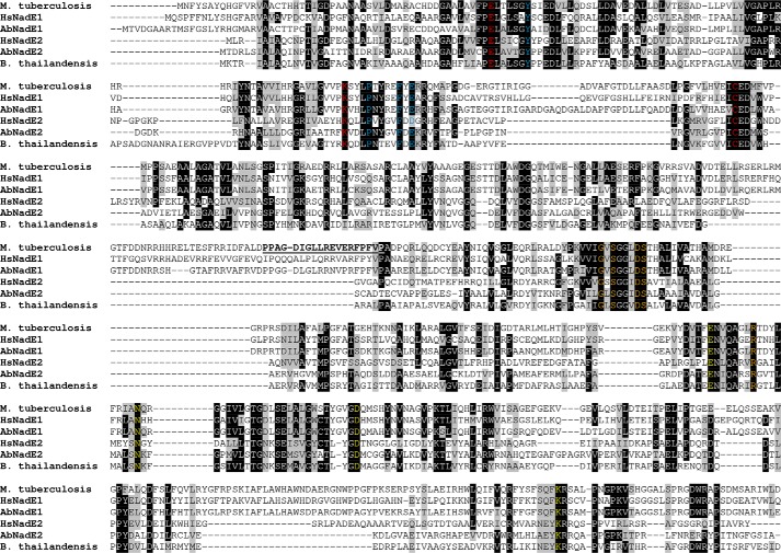
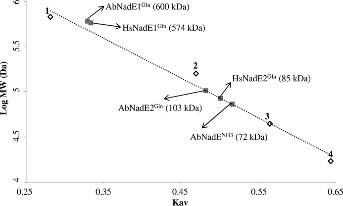
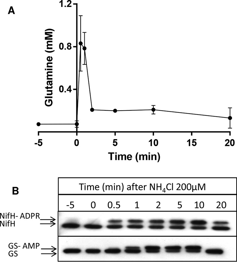
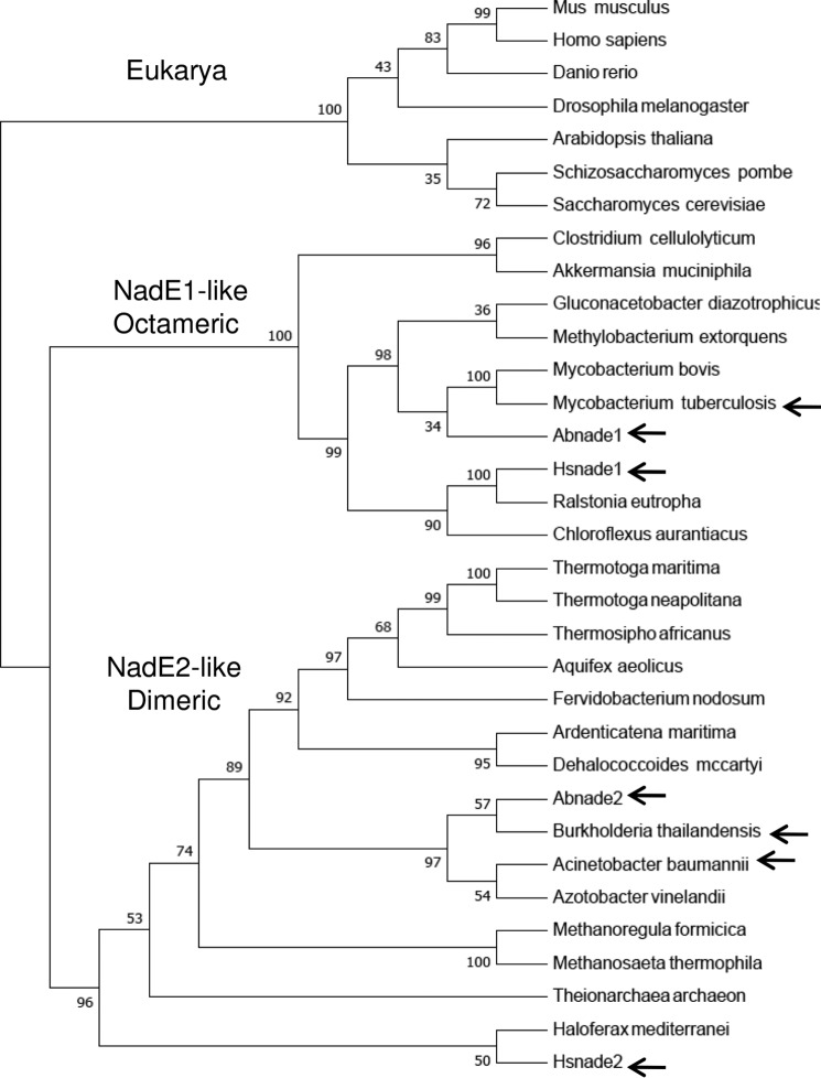
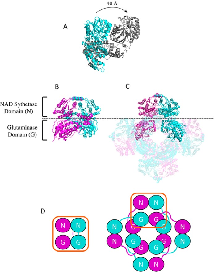
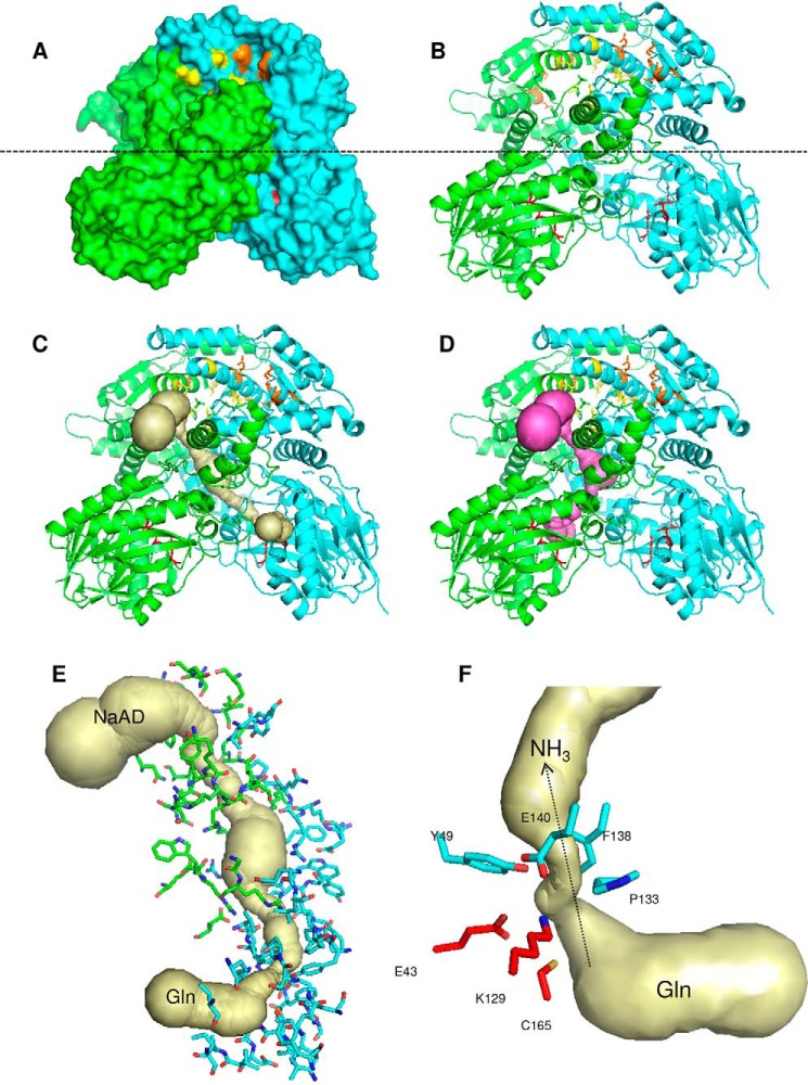
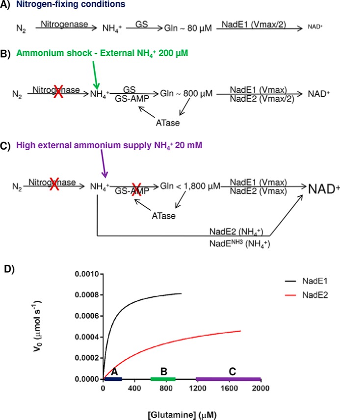
NADH (NAD+) and its reduced form NADH serve as cofactors for a variety of oxidoreductases that participate in many metabolic pathways. NAD+ also is used as substrate by ADP-ribosyl transferases and by sirtuins. NAD+ biosynthesis is one of the most fundamental biochemical pathways in nature, and the ubiquitous NAD+ synthetase (NadE) catalyzes the final step in this biosynthetic route. Two different classes of NadE have been described to date: dimeric single-domain ammonium-dependent NadENH3 and octameric glutamine-dependent NadEGln, and the presence of multiple NadE isoforms is relatively common in prokaryotes. Here, we identified a novel dimeric group of NadEGln in bacteria. Substrate preferences and structural analyses suggested that dimeric NadEGln enzymes may constitute evolutionary intermediates between dimeric NadENH3 and octameric NadEGln The characterization of additional NadE isoforms in the diazotrophic bacterium Azospirillum brasilense along with the determination of intracellular glutamine levels in response to an ammonium shock led us to propose a model in which these different NadE isoforms became active accordingly to the availability of nitrogen. These data may explain the selective pressures that support the coexistence of multiple isoforms of NadE in some prokaryotes.

Keywords: NAD biosynthesis; NadE; ammonia; ammonia assimilation; glutamine; glutamine synthase; nitrogen metabolism; nitrogenase.


Conflict of interest statement

The authors declare that they have no conflicts of interest with the contents of this article

Figures

Figure 1.
Figure 1.
Sequence alignment of different NadEGln. The sequences of M. tuberculosis, H. seropedicae NadE1Gln and NadE2Gln, A. brasilense NadE1Gln and NadE2Gln, and B. thailandensis were aligned using Clustal W. The threshold for shading was set as 60%. The conserved catalytic triad EKC present in the glutaminase domain is indicated in red. Conserved residues involved in NaAD and ATP binding are indicated in yellow and orange, respectively. Residues forming the ammonia tunnel constriction are indicated in blue. The interdomain loop in MtNadE1Gln is underlined.
Figure 2.
Figure 2.
Gel filtration analysis of the different NadE. Gel filtration was performed on a Superose 6 column (GE Healthcare), which was calibrated with a range of molecular mass standards (Bio-Rad): point 1, thyroglobulin; point 2, bovine γ-globulin; point 3, chicken ovalbumin; and point 4, equine myoglobin. The arrows indicate the estimated molecular weight of each NadE protein.
Figure 3.
Figure 3.
Intracellular levels of glutamine and NifH and GS modification after an ammonium shock in A. brasilense. A, average intracellular glutamine levels from triplicate experiments ± S.D. B, Western blotting analysis showing of the reversible post-translational modification of NifH and GS. The upper bands represent the ADP-ribosylated NifH (NifH-ADPR) or adenylylated GS (GS-AMP).
Figure 4.
Figure 4.
Phylogenetic analysis of selected NadEGln. The sequence of NadEGln was retrieved from NCBI and aligned using Clustal W. The phylogentic three was constructed by Neighbor-joining using MEGA 7. All positions containing gaps and missing data were eliminated from the data set. Bootstrap values were adjusted to 1000 replicates. Three groups were observed: 1) eukaryotic representatives; 2) NadE1-like octameric NadEGln; and 3) NadE2-like dimeric NadEGln. The relevant proteins with experimentally determined quaternary structure are indicated by arrows. A. baumannii, WP_065718975.1; Akkermansia muciniphila ATCC BAA-835, ACD04457.1; Aquifex aeolicus, NP_213654.1; Arabidopsis thaliana, NP_175906.1; Ardenticatena maritima, KPL88222.1; A. brasilense, AIB10872.1; A. brasilense, AIB14429.1; Azotobacter vinelandii DJ, YP_002798395.1; B. thailandensis, AOJ56104.1; Chloroflexus aurantiacus, ABY34602.1; Clostridium cellulolyticum H10, YP_002505537.1; Danio rerio, NP_001092723.1; Dehalococcoides mccartyi 195, YP_181837.1; Drosophila melanogaster, NP_572913.1; Fervidobacterium nodosum Rt17-B1, YP_001410277.1; Gluconacetobacter diazotrophicus PA1 5, YP_001601200.1; Haloferax mediterranei, AHZ23047.1; H. seropedicae, AKN65438.1; H. seropedicae, AKN67808.1; Homo sapiens, NP_060631.2; Methanoregula formicica, AGB01500.1; Methanosaeta thermophila PT, YP_843157.1; Methylobacterium extorquens CM4, YP_002423109.1; Mus musculus, NP_084497.1; Mycobacterium bovis AF2122/97, NP_856111.1; Ralstonia eutropha H16, YP_725265.1; S. cerevisiae S288C, NP_011941.1; Schizosaccharomyces pombe 972h-, NP_587771.1; Theionarchaea archaeon, KYK35595.1; Thermosipho africanus TCF52B, YP_002335497.1; T. maritima, AKE27162.1; Thermotoga neapolitana DSM 4359, YP_002534863.1; and M. tuberculosis, AMP30329.1.
Figure 5.
Figure 5.
Comparison between the structures of dimeric and octameric NadEGln. A, superimposition of the single chain of NadE2Gln from B. thailandensis (cyan) and NadE1Gln from M. tuberculosis (gray). The PDB structures 4FH4 and 3DLA were superimposed using COOT and visualized using PyMOL. Structures are colored by chain. B and C, comparison between the organization of dimeric NadEGln from B. thailandensis (B) and octameric NadEGln from M. tuberculosis (C). Only one dimer of the octameric M. tuberculosis NadE is shown without transparency for comparison with B. thailandensis. D, schematic organization of the NAD synthetase domain N and the glutaminase domain G in the dimeric and in the octameric conformation of NadEGln. Note that the relative orientations between the N and G domains remain the same though through a domain-swap interaction in the octameric case. The PDB structures 4FH4 and 3DLA were visualized using PyMOL.
Figure 6.
Figure 6.
Structure of the dimeric NadE2Gln from B. thailandensis. A and B, surface (A) and cartoon (B) representations; the monomers are colored green and cyan. The glutaminase catalytic triade residues are in red sticks. Residues involved in NaAD and ATP binding are shown as yellow and orange sticks, respectively. The dashed line indicates the separation of the NAD synthetase domain on the top and the glutaminase domain on the bottom. C and D, the ammonia tunnel connecting the glutaminase and NaAD synthetase domain in different protomers (C) and within the same protomer (D). The detailed structure of the intersubunit ammonia tunnel of the dimeric NadE2Gln from B. thailandensis is shown. E, residues forming the intersubunit ammonia tunnel from different promoters are indicated as green and cyan sticks, respectively. F, conserved residues forming the major constriction within the ammonia tunnel are show in cyan, and the glutaminase catalytic residues are in red. The figures were generated using PyMOL and the PDB entry 4F4H. The ammonia tunnel was identified by CAVER.
Figure 7.
Figure 7.
Model for the role of the three NadE in A. brasilense. A, under nitrogen-fixing conditions nitrogenase feeds ammonium into GS, and intracellular levels of glutamine are low (∼80 μm). NadE1Gln operates at Vmax/2 and promptly responds to small fluctuations in glutamine availability within this range (see the blue bar with Michaelis–Menten curves in D). NadE2Gln and NadENH3 activities are negligible leading to slow production of NAD+. B, upon an ammonium shock of NH4+ 200 μm, the intracellular glutamine rises to ∼800 μm, leading to total inhibition of nitrogenase by ADP-ribosylation and nearly full inhibition of GS by adenylylation. NadE1Gln operates at Vmax, whereas NadE2Gln operates at Vmax/2 and promptly responds to small fluctuations in glutamine availability within this range (see the green bar with Michaelis–Menten curves in D). NAD+ production is elevated. C, when external levels of ammonium are high, the high intracellular glutamine will lead to NadE1Gln and NadE2Gln to operate at Vmax using glutamine as substrate. Furthermore, because GS is fully inactive due adenylylation, free intracellular ammonium could be directly assimilated into NAD+ by NadE2Gln and presumably also by NadENH3 bypassing GS activity. The concert action of all three isoforms of NadE is likely to further enhance NAD+ production. D, velocities of the different A. brasilense NadE isoforms accordingly to the intracellular glutamine levels.

References

    1. Li J., Bonkowski M. S., Moniot S., Zhang D., Hubbard B. P., Ling A. J., Rajman L. A., Qin B., Lou Z., Gorbunova V., Aravind L., Steegborn C., and Sinclair D. A. (2017) A conserved NAD+ binding pocket that regulates protein–protein interactions during aging. Science 355, 1312–1317 10.1126/science.aad8242 - DOI - PMC - PubMed
    1. Moure V. R., Costa F. F., Cruz L. M., Pedrosa F. O., Souza E. M., Li X. D., Winkler F., and Huergo L. F. (2015) Regulation of nitrogenase by reversible mono-ADP-ribosylation. Curr. Top. Microbiol. Immunol. 384, 89–106 - PubMed
    1. Aravind L., Zhang D., de Souza R. F., Anand S., and Iyer L. M. (2015) The natural history of ADP-ribosyltransferases and the ADP-ribosylation system. Curr. Top. Microbiol. Immunol. 384, 3–32 - PMC - PubMed
    1. Bheda P., Jing H., Wolberger C., and Lin H. (2016) The substrate specificity of sirtuins. Annu. Rev. Biochem. 85, 405–429 10.1146/annurev-biochem-060815-014537 - DOI - PubMed
    1. Gazzaniga F., Stebbins R., Chang S. Z., McPeek M. A., and Brenner C. (2009) Microbial NAD metabolism: lessons from comparative genomics. Microbiol. Mol. Biol. Rev. 73, 529–541 10.1128/MMBR.00042-08 - DOI - PMC - PubMed

Publication types

MeSH terms

LinkOut - more resources