. 2018 Apr 10;115(15):3912-3917.
doi: 10.1073/pnas.1801524115. Epub 2018 Mar 26.

Anti-CTLA-4 therapy requires an Fc domain for efficacy

Affiliations

Anti-CTLA-4 therapy requires an Fc domain for efficacy

Jessica R Ingram et al. Proc Natl Acad Sci U S A. .

Abstract

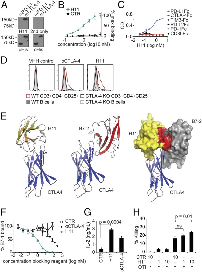
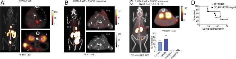
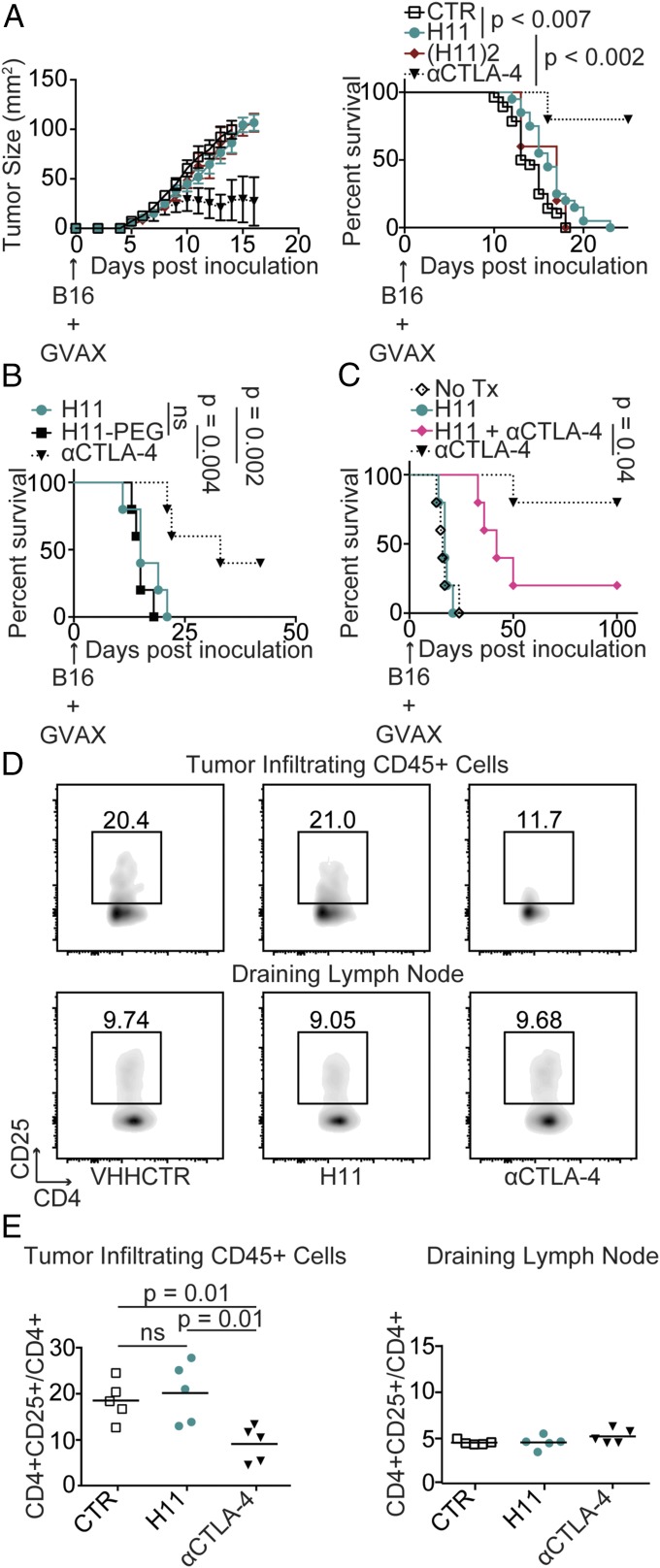
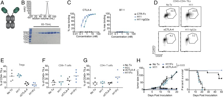
Ipilimumab, a monoclonal antibody that recognizes cytotoxic T lymphocyte antigen (CTLA)-4, was the first approved "checkpoint"-blocking anticancer therapy. In mouse tumor models, the response to antibodies against CTLA-4 depends entirely on expression of the Fcγ receptor (FcγR), which may facilitate antibody-dependent cellular phagocytosis, but the contribution of simple CTLA-4 blockade remains unknown. To understand the role of CTLA-4 blockade in the complete absence of Fc-dependent functions, we developed H11, a high-affinity alpaca heavy chain-only antibody fragment (VHH) against CTLA-4. The VHH H11 lacks an Fc portion, binds monovalently to CTLA-4, and inhibits interactions between CTLA-4 and its ligand by occluding the ligand-binding motif on CTLA-4 as shown crystallographically. We used H11 to visualize CTLA-4 expression in vivo using whole-animal immuno-PET, finding that surface-accessible CTLA-4 is largely confined to the tumor microenvironment. Despite this, H11-mediated CTLA-4 blockade has minimal effects on antitumor responses. Installation of the murine IgG2a constant region on H11 dramatically enhances its antitumor response. Coadministration of the monovalent H11 VHH blocks the efficacy of a full-sized therapeutic antibody. We were thus able to demonstrate that CTLA-4-binding antibodies require an Fc domain for antitumor effect.

Keywords: CTLA-4; cancer; checkpoint blockade; immunotherapy; single-domain antibody.


Conflict of interest statement

The authors declare no conflict of interest.

Figures

Fig. 1.
Fig. 1.
H11 recognizes CTLA-4. (A, Top) H11 immunoblot against His-tagged murine PD-L1 (mPD-L1) and mCTLA-4. “2nd only” refers to staining with secondary antibody alone. (A, Bottom) Corresponding anti-His immunoblots as loading controls. (B and C) Biotinylated H11 or VHH control (CTR) were incubated with plate-bound mCTLA-4-Fc (B) or recombinant Fc fusion proteins as indicated (C). Binding was detected by using streptavidin-HRP and tetramethylbenzidine (TMB). Data are normalized to the maximum signal (B) or represented as OD. Error bars show SD. (D) Flow cytometry on splenic populations as indicated from WT and CTLA-4 (inducibly) deleted (KO) mice using H11 or αCTLA-4 antibody as indicated. (E, Left) Ribbon drawing of the CTLA-4:H11 complex (PDB ID code 5ESM). CTLA-4 is in blue; H11 is in yellow. (E, Middle) Overall structure of CTLA-4:B7-2 complex (PDB ID code 1I85). CTLA-4 is in blue; B7-2 is in red. (E, Right) Surface rendering of CTLA-4 binding partners highlighting the steric clash between H11 and B7-2 binding sites (residues from H11 within 4.5-Å distance of B7-2, and vice versa, are colored red). (F) B7-1-Fc was incubated with plate-bound CTLA-4-Fc in the presence of H11, αCTLA-4, or VHH control (CTR). Binding was detected by using a biotinylated polyclonal antibody against B7-1 and streptavidin-HRP developed with TMB. Error bars show SD. (G) CD4+ T cells were isolated from the spleen by positive selection using magnetic beads and stimulated with plate-bound αCD3 and B7-1-Fc for 96 h in the presence of VHH or antibody as indicated. Secreted IL-2 levels were measured by ELISA. (H) Activated OT-I cells were cultured with IFN-γ–treated B16-ova cells in the presence of H11 or control VHH at the indicated concentrations. Cell viability was determined using CellTiterGlo and normalized to untreated cells. (G and H) Error bars show SEM. AD and FH represent at least three independent experiments. ns, nonsignificant.
Fig. 2.
Fig. 2.
In vivo imaging of CTLA-4 distribution by immuno-PET. PET-CT images of a naïve C57BL/6 WT mouse (A) or a mouse bearing a B16F10 melanoma (B) imaged with 18F-H11. (A and B, Left) Three-dimensional projection images overlaying the PET signal and the corresponding CT. (Upper Right) Transverse PET-CT overlays. (A, Bottom Right) Transverse PET-CT overlay of the abdomen capturing the organs of elimination: kidney (kd), intestines (int), gallbladder (gb), and urinary bladder (bl). (B, Lower Right) Transverse CT only. (A and B) Images are all window-leveled to the same intensity. (C) C57BL/6 WT mouse inoculated with B16 melanoma and treated with a combination of a tumor vaccine (GVAX) and anti-CTLA-4 (clone 9H10). After 10 d of treatment, mice were imaged with 89Zr-H11-PEG. The tumor (B16) and the vaccination site (GVAX) are marked. Three-dimensional projection image (Left) and transverse PET-CT overlays (Top Right) as in A and B. (Bottom Right) Standardized uptake values (SUVs) for 89Zr-H11-PEG imaged mice for tumor, GVAX, and two different muscle sites normalized to the average muscle signal. (D) Survival curve for mice imaged with 89Zr-H11-PEG compared with 89Zr-VHH-PEG control (n = 10). Results represent at least two independent experiments.
Fig. 3.
Fig. 3.
H11 has minimal antitumor activity in vivo. (A) C57BL/6 mice were inoculated with 5 × 10⁵ B16 cells and vaccinated with 5 × 10⁵ GM-CSF–secreting B16 cells (GVAX) on day 0. Mice were treated daily with 200 μg VHH control (CTR), H11, or H11 dimer [(H11)2] or every other day with 200 μg anti–CTLA-4 (αCTLA-4) antibodies, clone 9H10. (Left) Tumor size as measured by precision calipers. Error bars show SEM. Curves terminated when >50% of the group had been euthanized. (Right) Survival curve comparing treatment groups. Animals were euthanized when tumor reached 125 mm2. Control (n = 28), H11 (n = 20), H112 (n = 5), αCTLA-4 (n = 5). (B and C) Survival curves for mice inoculated with B16 and vaccinated as in A on day 0. (B) Mice were treated with 200 μg H11 daily or three times weekly with H11PEG or αCTLA-4 given at equimolar doses. (C) Starting on day 1, mice were left untreated or were treated daily with 200 μg H11, three times weekly with 200 μg αCTLA-4, or with a combination of the two treatments. (B and C) n = 5 for all groups. Results represent at least two independent experiments. (D and E) Mice were inoculated with B16 and vaccinated as in A on day 0. Mice were treated daily with VHH control (CTR) or H11 or three times weekly with αCTLA-4 antibodies starting on day 1. On day 10, mice were euthanized and tumor-infiltrating leukocytes (TILs) were isolated from resected tumors, or lymphocytes were isolated from the draining lymph node and analyzed by flow cytometry using the indicated antibodies. (D) Flow cytometry plots. (E) Quantification of data from D including multiple animals. ns, nonsignificant.
Fig. 4.
Fig. 4.
H11 fusion to IgG2a restores therapeutic efficacy. (A) Schematic representation of H11–IgG2a. (B) H11–IgG2a size-exclusion chromatogram (Top) and Coomassie-stained SDS/PAGE (Bottom). (C) Biotinylated H11, H11–IgG2a, or VHH-Fc control (CTR-Fc) were incubated with plate-bound mCTLA-4-Fc (Left) or mB7-1-Fc (Right). Binding was detected and analyzed as in Fig. 1B. (DG) Mice were inoculated with B16 and vaccinated as in Fig. 3A on day 0. Mice were treated daily with 100 µg H11 or twice weekly with 100 µg H11–IgG2a or 200 µg αCTLA-4 antibodies or were left untreated, starting on day 1. On day 11, mice were euthanized and TILs were isolated from resected tumors and analyzed by flow cytometry. (D) Flow-cytometry plots using the indicated antibodies. (E) Quantification of data from D including multiple animals. (F and G) Quantification of CD8+ TILs (F) and CD4+ TILs (G). (H) Mice were inoculated with B16, vaccinated, and treated as in DG. (Left) Tumor growth curves; (Right) Overall survival (*P < 0.05). Results represent at least two independent experiments. Tx, treatment.

References

    1. Baumeister SH, Freeman GJ, Dranoff G, Sharpe AH. Coinhibitory pathways in immunotherapy for cancer. Annu Rev Immunol. 2016;34:539–573. - PubMed
    1. Larkin J, et al. Combined nivolumab and ipilimumab or monotherapy in untreated melanoma. N Engl J Med. 2015;373:23–34. - PMC - PubMed
    1. Pardoll DM. The blockade of immune checkpoints in cancer immunotherapy. Nat Rev Cancer. 2012;12:252–264. - PMC - PubMed
    1. Hodi FS, et al. Improved survival with ipilimumab in patients with metastatic melanoma. N Engl J Med. 2010;363:711–723. - PMC - PubMed
    1. Robert C, et al. Ipilimumab plus dacarbazine for previously untreated metastatic melanoma. N Engl J Med. 2011;364:2517–2526. - PubMed

Publication types

MeSH terms