. 2018 Apr 10;115(15):3936-3941.
doi: 10.1073/pnas.1717338115. Epub 2018 Mar 26.

Reduction of lipid accumulation rescues Bietti's crystalline dystrophy phenotypes

Affiliations

Reduction of lipid accumulation rescues Bietti's crystalline dystrophy phenotypes

Masayuki Hata et al. Proc Natl Acad Sci U S A. .

Abstract

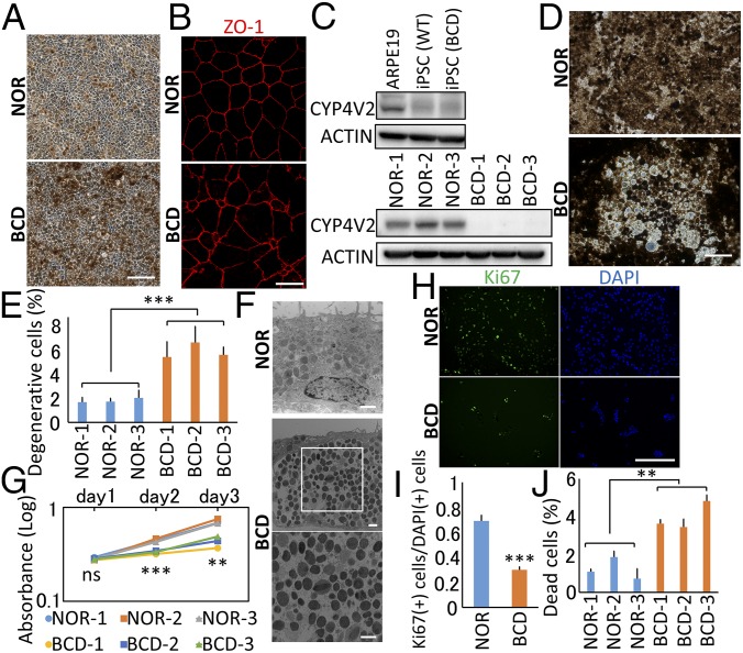
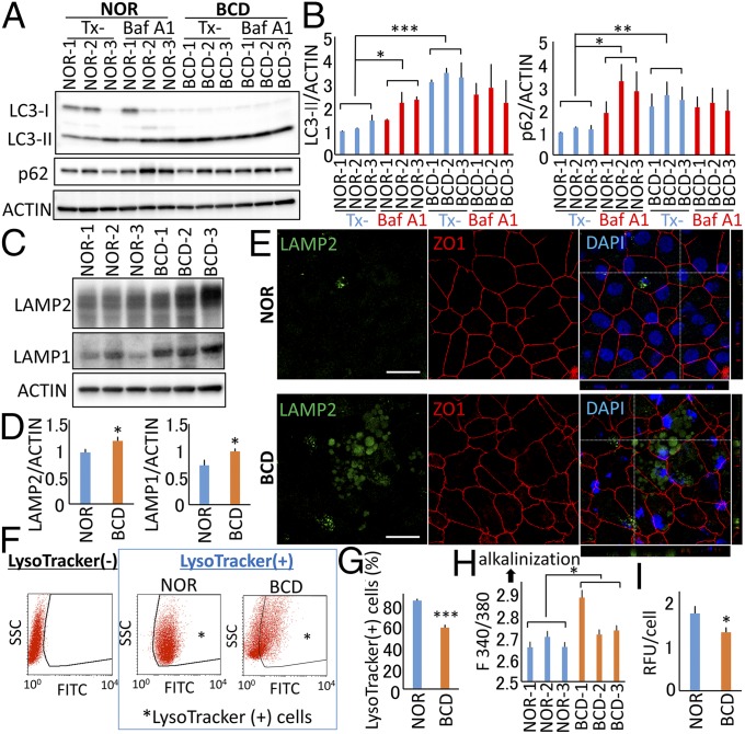
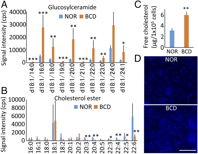
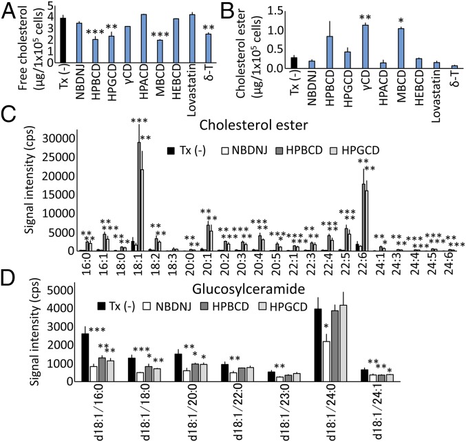
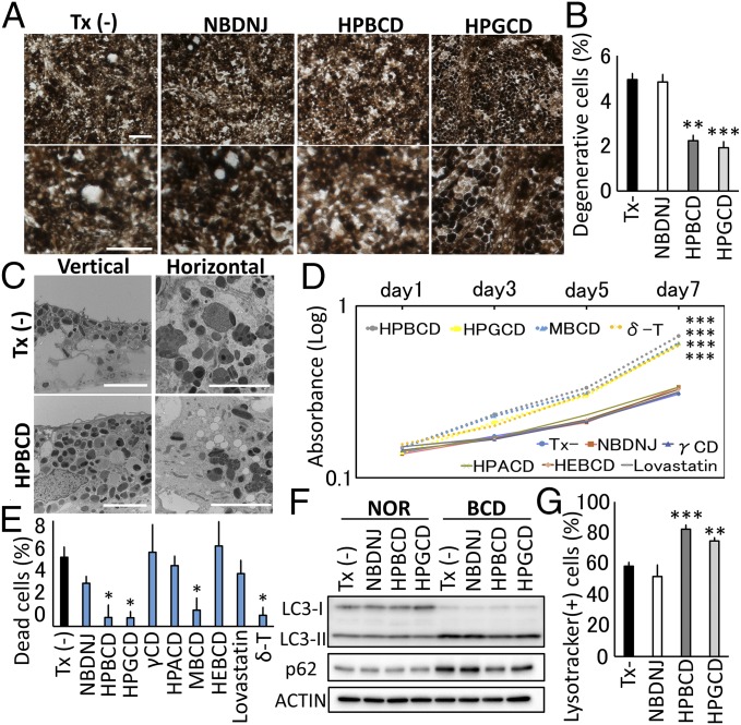
Bietti's crystalline dystrophy (BCD) is an intractable and progressive chorioretinal degenerative disease caused by mutations in the CYP4V2 gene, resulting in blindness in most patients. Although we and others have shown that retinal pigment epithelium (RPE) cells are primarily impaired in patients with BCD, the underlying mechanisms of RPE cell damage are still unclear because we lack access to appropriate disease models and to lesion-affected cells from patients with BCD. Here, we generated human RPE cells from induced pluripotent stem cells (iPSCs) derived from patients with BCD carrying a CYP4V2 mutation and successfully established an in vitro model of BCD, i.e., BCD patient-specific iPSC-RPE cells. In this model, RPE cells showed degenerative changes of vacuolated cytoplasm similar to those in postmortem specimens from patients with BCD. BCD iPSC-RPE cells exhibited lysosomal dysfunction and impairment of autophagy flux, followed by cell death. Lipidomic analyses revealed the accumulation of glucosylceramide and free cholesterol in BCD-affected cells. Notably, we found that reducing free cholesterol by cyclodextrins or δ-tocopherol in RPE cells rescued BCD phenotypes, whereas glucosylceramide reduction did not affect the BCD phenotype. Our data provide evidence that reducing intracellular free cholesterol may have therapeutic efficacy in patients with BCD.

Keywords: Bietti’s crystalline dystrophy; CYP4V2 gene; cholesterol; induced pluripotent stem cells; retinal pigment epithelium.


Conflict of interest statement

Conflict of interest statement: Kyoto University has applied for patents related to this study (JP2017/90296) with M.H. and H.O.I. listed as inventors.

Figures

Fig. 1.
Fig. 1.
Phenotypes of BCD patient-specific iPSC–RPE cells. (A) Bright-field micrographs taken after 3 mo of differentiation of BCD and NOR iPSCs into RPE cells (iPSC-RPE cells). (B) Immunocytochemical staining for ZO-1 in NOR and BCD iPSC-RPE cells. (C) Western blot analyses of CYP4V2 in a human RPE cell line (ARPE-19), NOR iPSCs, BCD iPSCs, NOR iPSC-RPE cells, and BCD iPSC-RPE cells. (D) Bright-field micrographs taken after 10 mo of differentiation of iPSCs into iPSC-RPE cells. (E) Evaluation of degenerative changes (having all the following features: vacuole formation, larger cell size, and pronounced pigmentation changes) in NOR and BCD iPSC-RPE cells. ***P < 0.001, BCD vs. NOR; Student’s t test; n = 3 in each group. (F) TEM of NOR and BCD iPSC-RPE cells cultured for 10 mo. (G) Evaluation of the proliferation rate using the reaction with water-soluble tetrazolium salts. **P = 0.003, ***P < 0.001, BCD vs. NOR; Student’s t test; n = 3 in each group. (H and I) Evaluation of the cell proliferation state using DAPI staining (blue) and immunocytochemical staining for Ki67 (green) in NOR and BCD iPSC-RPE progenitor cells. ***P < 0.001, BCD vs. NOR; Student’s t test; n = 4 in each group. (J) Evaluation of cell death. **P = 0.007, BCD vs. NOR; Student’s t test; n = 3 in each group. Error bars indicate SD. (Scale bars: 50 μm in A, D, and H; 10 μm in B; 1 μm in F.)
Fig. 2.
Fig. 2.
Autophagy and lysosome function in NOR and BCD iPSC-RPE cells. (A and B) Expression of LC3-II and p62 with or without bafilomycin-A1 (Baf A1, 20 nM) in NOR and BCD iPSC-RPE cells. NOR Tx− vs. NOR with Baf A1: *P = 0.0459 (LC3-II) and *P = 0.0496 (p62), paired t test, n = 3 in each group; NOR Tx− vs. BCD Tx−: ***P < 0.001 (LC3-II) and **P = 0.0013 (p62), Student’s t test, n = 3 in each group. (C and D) Expression of LAMP1 and LAMP2. *P < 0.05; Student’s t test; n = 3 derived from each of three lines. (E) Immunocytochemical staining for LAMP2 (green) and ZO1 (red). (Scale bars: 10 μm.) (F and G) FACS analysis of lysosome function using LysoTracker Green. ***P < 0.001, BCD vs. NOR; Student’s t test; n = 3 derived from each of three lines. (H) Lysosomal pH measurement using LysoSensor. The F340/380 ratio was determined. *P = 0.0495, BCD vs. NOR; Student’s t test; n = 3 in each group. (I) The cathepsin D activity levels measured using a fluorometric cathepsin D activity assay. *P = 0.032, BCD vs. NOR; Student’s t test; n = 3 derived from each of three lines. Error bars indicate SD.
Fig. 3.
Fig. 3.
Lipidomic analyses of NOR and BCD iPSC-RPE cells. (A and B) Untargeted lipidomics with LC-MS/MS in NOR and BCD iPSC-RPE cells. *P < 0.05, **P < 0.01, ***P < 0.001, NOR vs. BCD; Student’s t test; n = 10 and n = 8, respectively. (C) Free cholesterol concentration per cell number. **P = 0.005, NOR vs. BCD; Student’s t test; n = 3 derived from each of three lines. Error bars indicate SD. (D) Filipin staining of iPSC-RPE cells. (Scale bar, 50 μm.)
Fig. 4.
Fig. 4.
Effect of CDs on lipids in BCD iPSC-RPE cells. (A and B) Therapeutic effects of NBDNJ, cyclodextrins (CDs), lovastatin, and δ-T on intracellular free cholesterol (A) or cholesteryl ester levels (B) in BCD iPSC-RPE cells derived from three lines. ***P < 0.001, **P < 0.01, *P < 0.05 vs. no treatment [Tx(−)]; one-way ANOVA followed by the Dunnett’s test; n = 3. (C and D) LC-MS/MS–based lipidomic analyses of therapeutic effects of NBDNJ and CDs (HPBCD and HPGCD) on cholesteryl ester (C) or GlcCer (D). *P < 0.05, **P < 0.01, ***P < 0.001 vs. no treatment [Tx(−)]; one-way ANOVA followed by the Dunnett’s test; n = 3.
Fig. 5.
Fig. 5.
Therapeutic effect of free cholesterol reduction on phenotypes in BCD iPSC-RPE cells. (A) Bright-field micrographs of BCD iPSC-RPE cells treated with CDs (HPBCD and HPGCD) or NBDNJ. (B) Evaluation of degenerative changes in BCD iPSC-RPE cells treated with CDs (HPBCD and HPGCD) or NBDNJ. **P = 0.001, ***P < 0.001 vs. no treatment [Tx(−)]; one-way ANOVA followed by the Dunnett’s test; n = 3. (C) TEMs of BCD iPSC-RPE cells treated with HPBCD. (D) Evaluation of the proliferation rate in BCD iPSC-RPE progenitor cells treated with the indicated substances using the reaction with water-soluble tetrazolium salts. Day 7: ***P < 0.001 vs. no treatment [Tx(−)]; one-way ANOVA followed by Dunnett’s test; n = 4 in each group. (E) Evaluation of cell death in BCD iPSC-RPE progenitor cells treated with the indicated substances. *P < 0.05 vs. no treatment [Tx(–)]; one-way ANOVA followed by the Dunnett’s test; n = 3 in each group. (F) Therapeutic effects of the indicated substances on the abnormal autophagy parameters (LC3-II and p62) in BCD iPSC-RPE cells. (G) Therapeutic effects of the indicated substances on lysosome dysfunction in BCD iPSC-RPE cells using LysoTracker Green. ***P = 0.0007, **P = 0.0019 vs. no treatment [Tx(−)]; one-way ANOVA followed by Dunnett’s test; n = 3 derived from each of three lines. Error bars indicate SD. (Scale bars: 50 μm in A; 5 μm in C.)

Similar articles

Cited by

References

    1. Bietti GB. Uber familiares Vorkommen von “retinitis punctata albescens” (verbunden mit “dystrophia marginalis cristallinza cornea”), Glitzern des Glaskorpers und anderen degenerativen Augenverunderungen. Klin Monatsbl Augenheilkd. 1937;99:737.
    1. Mataftsi A, Zografos L, Millá E, Secrétan M, Munier FL. Bietti’s crystalline corneoretinal dystrophy: a cross-sectional study. Retina. 2004;24:416–426. - PubMed
    1. Li A, et al. Bietti crystalline corneoretinal dystrophy is caused by mutations in the novel gene CYP4V2. Am J Hum Genet. 2004;74:817–826. - PMC - PubMed
    1. Wilson DJ, Weleber RG, Klein ML, Welch RB, Green WR. Bietti’s crystalline dystrophy. A clinicopathologic correlative study. Arch Ophthalmol. 1989;107:213–221. - PubMed
    1. Halford S, et al. Detailed phenotypic and genotypic characterization of bietti crystalline dystrophy. Ophthalmology. 2014;121:1174–1184. - PubMed

Publication types

MeSH terms

Supplementary concepts