. 2018 Apr 10;115(15):3834-3839.
doi: 10.1073/pnas.1722336115. Epub 2018 Mar 26.

Sortase ligation enables homogeneous GPCR phosphorylation to reveal diversity in β-arrestin coupling

Affiliations

Sortase ligation enables homogeneous GPCR phosphorylation to reveal diversity in β-arrestin coupling

Dean P Staus et al. Proc Natl Acad Sci U S A. .

Abstract

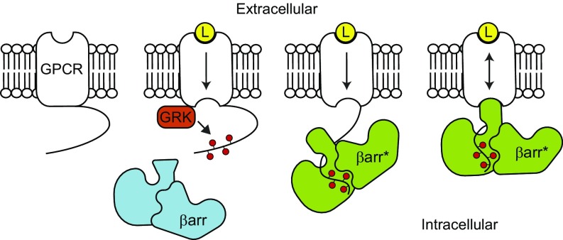
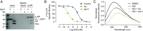
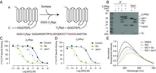
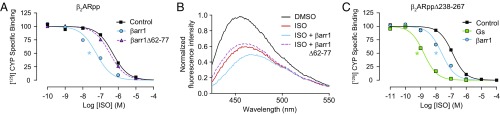
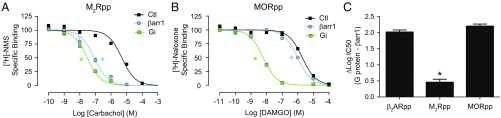
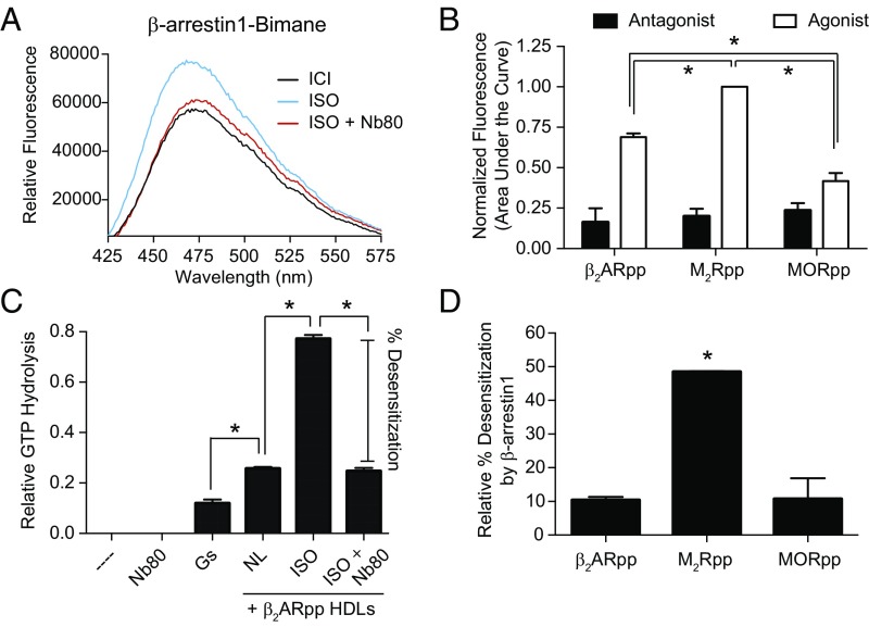
The ability of G protein-coupled receptors (GPCRs) to initiate complex cascades of cellular signaling is governed by the sequential coupling of three main transducer proteins, G protein, GPCR kinase (GRK), and β-arrestin. Mounting evidence indicates these transducers all have distinct conformational preferences and binding modes. However, interrogating each transducer's mechanism of interaction with GPCRs has been complicated by the interplay of transducer-mediated signaling events. For example, GRK-mediated receptor phosphorylation recruits and induces conformational changes in β-arrestin, which facilitates coupling to the GPCR transmembrane core. Here we compare the allosteric interactions of G proteins and β-arrestins with GPCRs' transmembrane cores by using the enzyme sortase to ligate a synthetic phosphorylated peptide onto the carboxyl terminus of three different receptors. Phosphopeptide ligation onto the β2-adrenergic receptor (β2AR) allows stabilization of a high-affinity receptor active state by β-arrestin1, permitting us to define elements in the β2AR and β-arrestin1 that contribute to the receptor transmembrane core interaction. Interestingly, ligation of the identical phosphopeptide onto the β2AR, the muscarinic acetylcholine receptor 2 and the μ-opioid receptor reveals that the ability of β-arrestin1 to enhance agonist binding relative to G protein differs substantially among receptors. Furthermore, strong allosteric coupling of β-arrestin1 correlates with its ability to attenuate, or "desensitize," G protein activation in vitro. Sortase ligation thus provides a versatile method to introduce complex, defined phosphorylation patterns into GPCRs, and analogous strategies could be applied to other classes of posttranslationally modified proteins. These homogeneously phosphorylated GPCRs provide an innovative means to systematically study receptor-transducer interactions.

Keywords: G protein-coupled receptor; allostery; phosphorylation; sortase; β-arrestin.


Conflict of interest statement

Conflict of interest statement: D.P.S., L.M.W., and R.J.L. are co-inventors on a patent application that covers methods described in this manuscript.

Figures

Fig. 1.
Fig. 1.
Illustration showing the two-step binding mode of β-arrestin. Ligand (L) binding to the extracellular orthosteric binding pocket leads to conformational changes within the GPCR transmembrane region to influence intracellular transducer binding. The phosphorylation (red circles) of the receptor C terminus by GPCR kinase (GRK) initiates the recruitment of β-arrestin (βarr). Conformational changes induced in βarr (βarr*) as a result of binding to the phosphorylated C terminus promotes coupling to the GPCR transmembrane core, which allosterically enhances ligand affinity.
Fig. 2.
Fig. 2.
Nonphosphorylated β2AR interacts with Gs heterotrimer but not β-arrestin1. (A) Coomassie-stained gel showing the coimmunoprecipitation of Gs heterotrimer (Gs) or β-arrestin1 (βarr1) with isoproterenol (ISO)-bound FLAG-β2AR. Loading controls represent 10% of input. (B) Competition binding experiments using radiolabeled [125I]-cyanopindolol (CYP). Gs increases ISO affinity for β2AR HDLs (log IC50: −8.88 ± 0.03) compared with no transducer (log IC50: −6.82 ± 0.03), but βarr1 does not (log IC50: −6.81 ± 0.02). Data shown are the mean of three independent experiments, with error bars representing SE. The green asterisk (*) indicates a log IC50 value significantly different from the control curve (P < 0.05, one-way ANOVA). (C) The fluorescence emission spectrum of bimane-labeled β2AR HDLs shows a rightward shift and decrease in fluorescence upon addition of ISO, indicative of receptor activation. The effects of ISO are enhanced by Gs but not βarr1. Data shown are representative of three independent experiments.
Fig. 3.
Fig. 3.
Sortase ligation of a phosphopeptide onto the β2AR restores its allosteric interaction with β-arrestin1. (A) Cartoon schematic of sortase ligation method. A synthetic phosphopeptide (pp) derived from the vasopressin-2-receptor (V2R) with three N-terminal glycine residues (GGG-V2Rpp) is ligated onto receptors containing a C-terminal LPETGGH recognition motif. In the sequence of GGG-V2Rpp below the schematic, phosphorylated residues are highlighted in red. (B) Coomassie-stained gel showing the coimmunoprecipitation of heterotrimeric Gs and β-arrestin1 (βarr1) with isoproterenol (ISO)-bound, phosphopeptide-ligated FLAG-β2AR (β2ARpp). Fab30 binds specifically to V2Rpp-bound βarr1 (10). Loading controls represent 10% of input. (C and D) Competition binding experiments using radiolabeled [125I]-cyanopindolol (CYP) with HDLs containing (C) β2ARpp or (D) β2AR ligated to a nonphosphorylated version of the V2R peptide (β2ARnp). Gs increases the affinity of ISO for both β2ARpp and β2ARnp HDLs (log IC50: −9.15 ± 0.03, −9.02 ± 0.04, respectively) compared with no transducer (log IC50: −6.24 ± 0.04, −6.42 ± 0.09, respectively), but βarr1 only increases ISO affinity for β2ARpp HDLs (log IC50: −7.14 ± 0.07) and not β2ARnp HDLs (log IC50: −6.49 ± 0.04). Data shown in C and D are the mean of at least three independent experiments, with error bars representing SE, and asterisks (*) indicate a log IC50 value significantly different from the control curve (P < 0.05, one-way ANOVA). (E) The effects of ISO on the HDL-β2ARpp-bimane fluorescence emission spectrum are enhanced by Gs and βarr1. Data shown are representative of three independent experiments.
Fig. 4.
Fig. 4.
The allosteric interaction between phosphorylated β2AR and β-arrestin1 requires the finger loop of β-arrestin1 but does not require the third intracellular loop of the β2AR. (A) In competition radioligand binding with β2ARpp HDLs as described in Fig. 3C, a finger loop deletion mutant of β-arrestin1 (βarr1) (Δ62–77) has minimal effect on isoproterenol (ISO) binding (log IC50s: no transducer, −6.30 ± 0.05; βarr1, −7.25 ± 0.07; βarr1 Δ62–77, −6.48 ± 0.03). (B) βarr1Δ62–77 does not intensify the effects of ISO on the fluorescence spectrum of β2ARpp-bimane HDLs. Data shown are representative of three independent experiments. (C) In competition radioligand binding with β2ARpp HDLs containing a deletion of the third intracellular loop (Δ238–267), both Gs (log IC50: −8.85 ± 0.03) and βarr1 (log IC50: −7.63 ± 0.06) retain their ability to increase isoproterenol (ISO) affinity (no transducer, log IC50: −6.91 ± 0.04). Data shown in A and C are the mean of at least three independent experiments, with error bars representing SE, and asterisks (*) indicate a log IC50 value significantly different from the control curve (P < 0.05, one-way ANOVA).
Fig. 5.
Fig. 5.
The allosteric enhancement of agonist binding induced by β-arrestin1 varies among different receptors. (A) Competition binding experiments with sortase-ligated M2Rpp HDLs, using [3H]-N-methyl-scopolamine (NMS) as the tracer. Heterotrimeric Gi (100 nM, log IC50: −7.51 ± 0.06) and β-arrestin1 (βarr1) (1 μM, log IC50: −7.06 ± 0.08) increase the affinity of the agonist carbachol to a similar extent (no transducer, log IC50: −5.31 ± 0.09). (B) Competition binding experiments with sortase-ligated MORpp HDLs, using [3H]-naloxone as the tracer. Gi (1 μM, log IC50: −8.22 ± 0.05) increases the affinity of the agonist DAMGO to a far greater extent than βarr1 (1 μM, log IC50: −6.02 ± 0.05) (no transducer, log IC50: −5.71 ± 0.06). Data in A and B are the mean of three independent experiments, with error bars representing SE, and asterisks (*) indicate a log IC50 value significantly different from the control curve (P < 0.05, one-way ANOVA). (C) Comparison of the difference in agonists’ log IC50 values in the presence of their cognate G proteins versus βarr1 for sortase-ligated β2ARpp (Fig. 3C), M2Rpp (A), and MORpp (B) HDLs.
Fig. 6.
Fig. 6.
The extent to which β-arrestin1 engages receptors’ transmembrane cores varies among different receptors. (A) Fluorescence spectra of β-arrestin1 (βarr1) labeled with monobromobimane at residue 70 in the finger loop. Activation of HDL-β2ARpp by the agonist isoproterenol (ISO) increases βarr1-bimane fluorescence, which is blocked by Nb80 binding to the receptor TM core. Data shown are representative of three independent experiments. (B) Comparison of βarr1-bimane fluorescence by agonist activation of β2ARpp, M2Rpp, and MORpp HDLs. The area under the fluorescence emission spectra were determined and normalized to M2Rpp plus iperoxo (the maximum signal) in each experiment (see Fig. S4 for representative spectra). All three receptors are significantly different from one another (*P < 0.05), and β2ARpp and M2Rpp are significantly different from their respective antagonist controls (not indicated, P < 0.05). (C) An in vitro GTPase assay measuring GTP hydrolysis as a readout of G protein activation. The basal level of GTP hydrolysis induced by G protein is robustly increased by HDL-β2ARpp HDLs in the presence of ISO compared with no ligand (NL) (*P < 0.05), which is blocked (desensitization) by the addition of Nb80 (no significant difference between NL and ISO + Nb80). (D) Inhibition (percentage desensitization) of G protein activation by βarr1 is strongest at M2Rpp and significantly different from β2ARpp and MORpp HDLs (*P < 0.05) (see also Fig. S5). Data in BD are the mean of at least three independent experiments, with error bars representing SE; P values were determined by one-way ANOVA.

References

    1. Lagerström MC, Schiöth HB. Structural diversity of G protein-coupled receptors and significance for drug discovery. Nat Rev Drug Discov. 2008;7:339–357. - PubMed
    1. Manglik A, Kruse AC. Structural basis for G protein-coupled receptor activation. Biochemistry. 2017;56:5628–5634. - PMC - PubMed
    1. Lefkowitz RJ. A brief history of G-protein coupled receptors (Nobel Lecture) Angew Chem Int Ed Engl. 2013;52:6366–6378. - PubMed
    1. Neves SR, Ram PT, Iyengar R. G protein pathways. Science. 2002;296:1636–1639. - PubMed
    1. Benovic JL, et al. Functional desensitization of the isolated beta-adrenergic receptor by the beta-adrenergic receptor kinase: Potential role of an analog of the retinal protein arrestin (48-kDa protein) Proc Natl Acad Sci USA. 1987;84:8879–8882. - PMC - PubMed

Publication types

MeSH terms