. 2018 Apr 24;115(17):4405-4410.
doi: 10.1073/pnas.1719251115. Epub 2018 Mar 26.

Structural determinants and cellular environment define processed actin as the sole substrate of the N-terminal acetyltransferase NAA80

Affiliations

Structural determinants and cellular environment define processed actin as the sole substrate of the N-terminal acetyltransferase NAA80

Marianne Goris et al. Proc Natl Acad Sci U S A. .

Abstract

N-terminal (Nt) acetylation is a major protein modification catalyzed by N-terminal acetyltransferases (NATs). Methionine acidic N termini, including actin, are cotranslationally Nt acetylated by NatB in all eukaryotes, but animal actins containing acidic N termini, are additionally posttranslationally Nt acetylated by NAA80. Actin Nt acetylation was found to regulate cytoskeletal dynamics and motility, thus making NAA80 a potential target for cell migration regulation. In this work, we developed potent and selective bisubstrate inhibitors for NAA80 and determined the crystal structure of NAA80 in complex with such an inhibitor, revealing that NAA80 adopts a fold similar to other NAT enzymes but with a more open substrate binding region. Furthermore, in contrast to most other NATs, the substrate specificity of NAA80 is mainly derived through interactions between the enzyme and the acidic amino acids at positions 2 and 3 of the actin substrate and not residues 1 and 2. A yeast model revealed that ectopic expression of NAA80 in a strain lacking NatB activity partially restored Nt acetylation of NatB substrates, including yeast actin. Thus, NAA80 holds intrinsic capacity to posttranslationally Nt acetylate NatB-type substrates in vivo. In sum, the presence of a dominant cotranslational NatB in all eukaryotes, the specific posttranslational actin methionine removal in animals, and finally, the unique structural features of NAA80 leave only the processed actins as in vivo substrates of NAA80. Together, this study reveals the molecular and cellular basis of NAA80 Nt acetylation and provides a scaffold for development of inhibitors for the regulation of cytoskeletal properties.

Keywords: N-terminal acetylation; NAA80; acetyltransferase; actin; inhibitor.


Conflict of interest statement

The authors declare no conflict of interest.

Figures

Fig. 1.
Fig. 1.
NAA80 activity and inhibition. (A) Purified maltose-binding protein (MPB) tagged human NAA80 (MBP-HsNAA80) was tested for in vitro NAT activity toward a panel of potential substrates representing the different substrate classes (NatA–H). (B) Structures for the NAA80 bisubstrate inhibitors. (C) IC50 values for inhibitors measured in an Nt acetylation assay for NAA80. (D) Ki values for CoA-Ac-DDDI-NH2 for different human NATs measured in an Nt acetylation assay. All reactions were performed at least three times and in triplicate, and errors bars represent SD of each measurement.
Fig. 2.
Fig. 2.
Overall structure of the binary DmNAA80/CoA-Ac-DDDI-NH2 complex. (A) Cellular Nt acetylation of β- and γ-actin by DmNAA80. (B) Overall structure of DmNAA80 in green and bisubstrate inhibitor CoA-Ac-DDDI-NH2 (yellow). (C) Structural alignment of DmNAA80 and it substrate (green) with other NATs: SpNAA10 (PDB ID code 4KVM) and its substrate in gray (23), CaNAA20 (PDB ID code 5K18) and its substrate in raspberry (26), HsNAA50 (PDB ID code 3TFY) and its substrate in pink (24), and HsNAA60 (PDB ID code 5ICV) and its substrate in light blue (21). The shifted positioning of the DmNAA80 substrate is indicated with a black arrow. Also note the shifted α1–α2 loop of DmNAA80 compared with the other NATs.
Fig. 3.
Fig. 3.
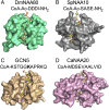
The substrate binding groove of DmNAA80. (A) DmNAA80 (green) bound to CoA-Ac-DDDI-NH2 (yellow) illustrating an open binding cleft. (B) SpNAA10 (PDB ID code 4KVM; gray) with CoA-Ac-SASE-NH2 (yellow) (23). (C) Lysine acetyltransferase GCN5 from Tetrahymena termophila (PDB ID code 1QSN; tan) with CoA-KSTGGKAPRKQ (yellow) (20); acetylated lysine is indicated in bold. (D) CaNAA20 (pink; PDB ID code 5K18) with CoA-Ac-MDSEVAALVID (yellow) (26).
Fig. 4.
Fig. 4.
DmNAA80 active site and peptide binding. DmNAA80 is shown in green, and the bisubstrate inhibitor CoA-Ac-DDDI-NH2 is shown as yellow stick. Bonds are shown as dotted black lines. Key secondary structure elements are labeled. (A) Electron density map (|Fo| − |Fc| omit map) contoured at 3.0σ of the peptide moiety of the bisubstrate inhibitor. (B) Electrostatic surface of the DmNAA80 substrate binding site as calculated using the program Delphi (34). Blue indicates positive charges, and red indicates negative charges. Charged residues are indicated in white. (C) Close-up view of interactions between the inhibitor and the DmNAA80 substrate binding site. (D) Close-up view of D1 of the inhibitor in the active site. A water molecule is shown as a red sphere as well as three of its potential H bonds. (E) Close-up view of D2 of the inhibitor in the active site. (F) Close-up view of D3 of the inhibitor in the active site. A water molecule is shown as a red sphere as well as two of its potential H bonds. (G) C. albicans NAA20 (CaNAA20; PDB ID code 5K18; raspberry) (26) and DmNAA80 superimposed with the first residue of their substrates (M1 and D1, respectively) in their binding pockets. NAA20 residues are labeled in raspberry.

Comment in

  • NATure of actin amino-terminal acetylation.
    Rubenstein PA, Wen KK. Rubenstein PA, et al. Proc Natl Acad Sci U S A. 2018 Apr 24;115(17):4314-4316. doi: 10.1073/pnas.1803804115. Epub 2018 Apr 9. Proc Natl Acad Sci U S A. 2018. PMID: 29632202 Free PMC article. No abstract available.

References

    1. Arnesen T, et al. Proteomics analyses reveal the evolutionary conservation and divergence of N-terminal acetyltransferases from yeast and humans. Proc Natl Acad Sci USA. 2009;106:8157–8162. - PMC - PubMed
    1. Bienvenut WV, et al. Comparative large scale characterization of plant versus mammal proteins reveals similar and Idiosyncratic N-α-Acetylation features. Mol Cell Proteomics. 2012;11:M111.015131. - PMC - PubMed
    1. Aksnes H, Drazic A, Marie M, Arnesen T. First things first: Vital protein marks by N-terminal acetyltransferases. Trends Biochem Sci. 2016;41:746–760. - PubMed
    1. Aksnes H, et al. An organellar nα-acetyltransferase, naa60, acetylates cytosolic N termini of transmembrane proteins and maintains Golgi integrity. Cell Rep. 2015;10:1362–1374. - PubMed
    1. Dinh TV, et al. Molecular identification and functional characterization of the first Nα-acetyltransferase in plastids by global acetylome profiling. Proteomics. 2015;15:2426–2435. - PMC - PubMed

Publication types

MeSH terms