. 2018 Jun;24(6):761-768.
doi: 10.1261/rna.065508.117. Epub 2018 Mar 26.

HnRNP L represses cryptic exons

Affiliations

HnRNP L represses cryptic exons

Sean P McClory et al. RNA. 2018 Jun.

Abstract

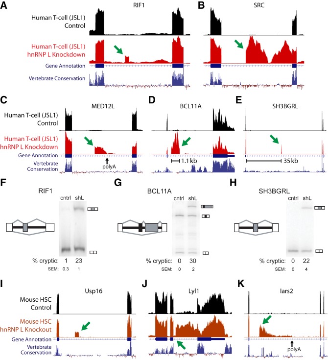
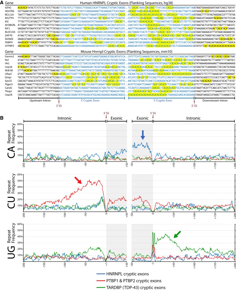
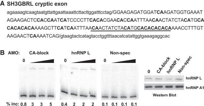
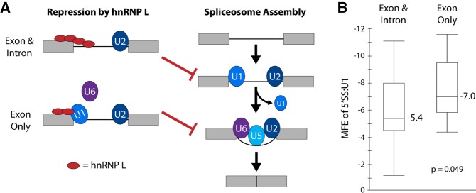
The fidelity of RNA splicing is regulated by a network of splicing enhancers and repressors, although the rules that govern this process are not yet fully understood. One mechanism that contributes to splicing fidelity is the repression of nonconserved cryptic exons by splicing factors that recognize dinucleotide repeats. We previously identified that TDP-43 and PTBP1/PTBP2 are capable of repressing cryptic exons utilizing UG and CU repeats, respectively. Here we demonstrate that hnRNP L (HNRNPL) also represses cryptic exons by utilizing exonic CA repeats, particularly near the 5'SS. We hypothesize that hnRNP L regulates CA repeat repression for both cryptic exon repression and developmental processes such as T cell differentiation.

Keywords: HNRNPL; HnRNP L; alternative splicing; cryptic exons; dinucleotide repeats.


Figures

FIGURE 1.
FIGURE 1.
Human and mouse hnRNP L cryptic exons viewed in the UCSC Genome Browser (hg38, mm10). Human cryptic cassette exon (A), cryptic exon extension (B), and cryptic 3′ end exon (C). Examples of variability: a large cassette exon (D) and a cassette exon flanked by long introns (E). (FH) Representative RTPCR gels showing splicing of hnRNP L repressed cryptic exons for both control JSL1 cells (cntrl) and JSL1 cells transfected with short-hairpin RNA against hnRNP L (shL). Splice graphs of each splicing event are shown to the left of each gel with the cryptic exon in gray, constitutive (upstream and downstream) exons in white, and intersecting alternative exons in black. The average percent inclusion of the cryptic exon (% cryptic) and standard error of the mean (SEM) for at least two replicates is shown (see Supplemental Fig. 1 for other examples). Mouse cryptic cassette exon (I), cryptic exon extension (J), and cryptic 3′ end exon (K). Genes displayed in UCSC views: RIF1 (A), SRC (B), MED12L (C), BCL11A (D), SH3BGRL (E), Usp16 (I), Lyl1 (J), and Iars2 (K).
FIGURE 2.
FIGURE 2.
Cryptic exon repressors demonstrate differential preference in repeat location. (A) Examples of sequences flanking hnRNP L cassette cryptic exons found in human (top) and mouse (bottom) data sets. Sequences are aligned to the 3′SS and 5′SS and CA repeat clusters are highlighted in yellow. (B) Frequency of CA, CU, and UG dinucleotide repeats (top, middle, bottom) for cassette cryptic exons associated with hnRNP L, PTBP1/PTBP2, and TDP-43 (blue, red, green). hnRNP L cryptic cassette exons have a high frequency of exonic CA repeats near the 5′SS. PTBP1 and PTBP2 cryptic cassette exons exhibit CU repeats mostly upstream of the 3′SS, with some also located in the exon. TDP-43 cryptic cassette exons primarily have UG repeats downstream from the 5′SS, although some repeats can also be upstream of the 3′SS.
FIGURE 3.
FIGURE 3.
Blocking CA repeats mimics depletion of hnRNP L in inclusion of the SH3BGRL cryptic exon. (A) Sequence of the SH3BGRL cryptic exon (uppercase) and flanking intron (lowercase) with sequence complementary to CA blocking AMO (underlined). CA dinucleotides are in bold. (B) RT-PCR gel showing inclusion of SH3BGRL cryptic exon upon transfection of CA blocking AMO, hnRNP L-depleting AMO or an unrelated GT-rich AMO. Western blot confirming depletion of hnRNP L upon treatment with the hnRNP L AMO, but not others, shown on right. HnRNP L depletion by AMO was performed as described previously (Cole et al. 2015). Translation blocking AMO for hnRNP L was used at 2, 5, and 10 nmol. CA and nonspecific AMO were used at 5, 10, and 20 nmol.
FIGURE 4.
FIGURE 4.
Model for different mechanisms of hnRNP L repression of weak and strong 5′ splice sites. (A) Schematic of normal splicesome assembly (right) and mechanisms by which hnRNP L represses normal assembly. Exons are designated as gray boxes, introns are black lines. The U snRNP components of the spliceosome are indicated by blue and purple ovals. HnRNP L is indicated by a red oval. HnRNP L can either block accessibility of the 5′SS to U1 (exon and intron), or inhibit exchange of U1 for U6 (exon only). In either case, assembly of the final spliceosomal complex is prevented. (B) Minimal free energy (MFE) of the association of the 5′SS with the U1 snRNA for hnRNP L-repressed exons that have CA dinucleotides in the exon and/or intron flanking the 5′SS. See Supplemental Table 1 for details. As described in the text, binding of hnRNP L to the exon only is predicted to repress U1–U6 exchange and is typically marked by a strong 5′SS:U1 interaction. P-value is a two-tailed t-test.

References

    1. Afgan E, Baker D, van den Beek M, Blankenberg D, Bouvier D, Čech M, Chilton J, Clements D, Coraor N, Eberhard C, et al. 2016. The Galaxy platform for accessible, reproducible and collaborative biomedical analyses: 2016 update. Nucleic Acids Res 44: W3–W10. - PMC - PubMed
    1. Ashiya M, Grabowski PJ. 1997. A neuron-specific splicing switch mediated by an array of pre-mRNA repressor sites: evidence of a regulatory role for the polypyrimidine tract binding protein and a brain-specific PTB counterpart. RNA 3: 996–1015. - PMC - PubMed
    1. Ayala YM, Pantano S, D'Ambrogio A, Buratti E, Brindisi A, Marchetti C, Romano M, Baralle FE. 2005. Human, Drosophila, and C. elegans TDP43: nucleic acid binding properties and splicing regulatory function. J Mol Biol 348: 575–588. - PubMed
    1. Blatter M, Dunin-Horkawicz S, Grishina I, Maris C, Thore S, Maier T, Bindereif A, Bujnicki JM, Allain FHT. 2015. The signature of the five-stranded vRRM fold defined by functional, structural and computational analysis of the hnRNP L protein. J Mol Biol 427: 3001–3022. - PubMed
    1. Boutz PL, Stoilov P, Li Q, Lin C, Chawla G, Ostrow K, Shiue L, Ares M Jr, Black DL. 2007. A post-transcriptional regulatory switch in polypyrimidine tract-binding proteins reprograms alternative splicing in developing neurons. Genes Dev 21: 1636–1652. - PMC - PubMed

Publication types

Substances

LinkOut - more resources