

doi: 10.1038/s41598-018-23408-0.
Modeling the effects of lipid peroxidation during ferroptosis on membrane properties
Affiliations
- PMID: 29581451
- PMCID: PMC5979948
- DOI: 10.1038/s41598-018-23408-0
Item in Clipboard
Modeling the effects of lipid peroxidation during ferroptosis on membrane properties
Sci Rep.
.
Abstract
Ferroptosis is a form of regulated cell death characterized by the accumulation of lipid hydroperoxides. There has been significant research on the pathways leading to the accumulation of oxidized lipids, but the downstream effects and how lipid peroxides cause cell death during ferroptosis remain a major puzzle. We evaluated key features of ferroptosis in newly developed molecular dynamics models of lipid membranes to investigate the biophysical consequences of lipid peroxidation, and generated hypotheses about how lipid peroxides contribute to cell death during ferroptosis.
Conflict of interest statement
The authors declare no competing interests.
Figures
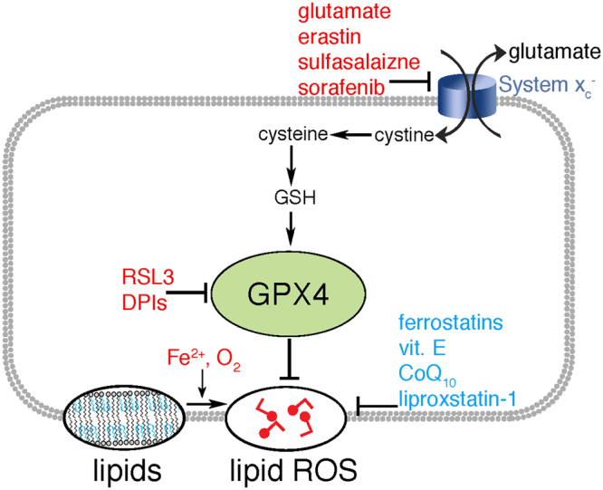
Inducers (red), and inhibitors (blue) of ferroptosis. GPX4 protects cells from lipid peroxidation; its inhibition by the depletion of GSH, or more directly through its binding with molecules such as RSL3, triggers accumulation of lipid oxygen reactive species (ROS), and trigger cell death.

The role of lipids in ferroptosis. Polyunsaturated fatty acids (PUFAs), depicted in light blue, are acetylated by acyl-CoA synthetase and inserted into phospholipids with saturated fatty acids (SFAs). Ferroptosis involves oxidation (red circles) of PUFAs, which can be repaired with GPX4 by reducing the ox-PUFAs to lipid alcohols (blue triangles). In the absence of GPX4, ox-PUFAs can result the release of reactive oxygen species (red fragment) and membrane destruction.

Peroxidation and coarse graining. (A) Peroxidation at double bond. This only happens at bis-allylic carbons. (B) Different MARTINI beads for three carbons with a double bond (blue), three saturated carbons (purple), and a peroxide (red). (C) A non-oxidized coarse-grained lipid. (D) An oxidized coarse-grained lipid.
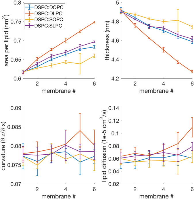
The SFA:PUFA experiment consists of 4 different scans; DSPC:DOPC, DSPC:DLPC, DSPC:SOPC, and DSPC:SLPC. Each one of these shows increasing area per lipid and decreasing thickness as it transitions towards a ferroptosis-relevant lipid. Lipid diffusion also increases slightly, with a more pronounced effect with DLPC.

The lipid:ox-lipid experiment consists of 4 scans; DLPE:oxDLPE, SLPE:oxSLPE, DHPE:ox1DHPE, and the more highly oxidized DHPE:ox2DHPE. These reveal increased area per lipid, decreased thickness, and increased curvature.
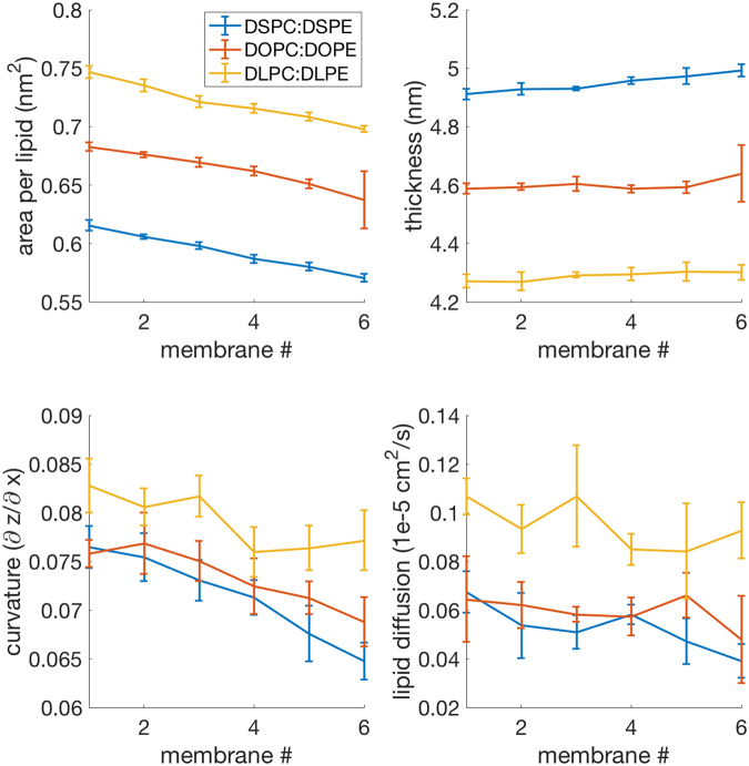
The PC:PE experiment consists of 3 scans; DSPC:DSPE, DOPC:DOPE, DLPC:DLPE. These reveal slight decreases in area per lipid, slight increases in thickness, curvature is reduced, and lipid diffusion remains constant.

A cross-section of four membranes, highlighting the changes caused by ferroptosis. (A) A membrane composed of 100% DSPC. (B) A membrane composed of 100% oxidized DLPC. (C) A membrane composed of 100% oxDLPE. (D) A membrane composed of 100% ox2DHPE. Peroxide beads are shown in red, double-bond beads are shown in purple, saturated carbon beads are shown in cyan, glycerol linkers are pink, and the head groups are blue and yellow.

Experimental results demonstrating altered membrane properties following lipid peroxidation. (A) DOPC and fluorescent lipid C5-Bodipy-HPC. (B) A vesicle with 95% DOPC and 5% Bodipy. Upon illumination in panel (b), photobleaching begins and lipid peroxides form. Within 10 seconds, from panels (b) to (e), the membrane visibly fluctuates more and more and highly curved area appear. (a and f) The same GUV observed before and after illumination by phase contrast microscopy. Scale bar, 10 μm. (C) Relative area change versus time as measured with micropipette aspiration (N = 10). t = 0 corresponds to the start of the fluorescent illumination.
References
Publication types
MeSH terms
Substances
Grants and funding
LinkOut - more resources
Full Text Sources
Other Literature Sources
