. 2018 Mar 26;9(1):1228.
doi: 10.1038/s41467-018-03566-5.

Microglia remodel synapses by presynaptic trogocytosis and spine head filopodia induction

Affiliations

Microglia remodel synapses by presynaptic trogocytosis and spine head filopodia induction

Laetitia Weinhard et al. Nat Commun. .

Abstract

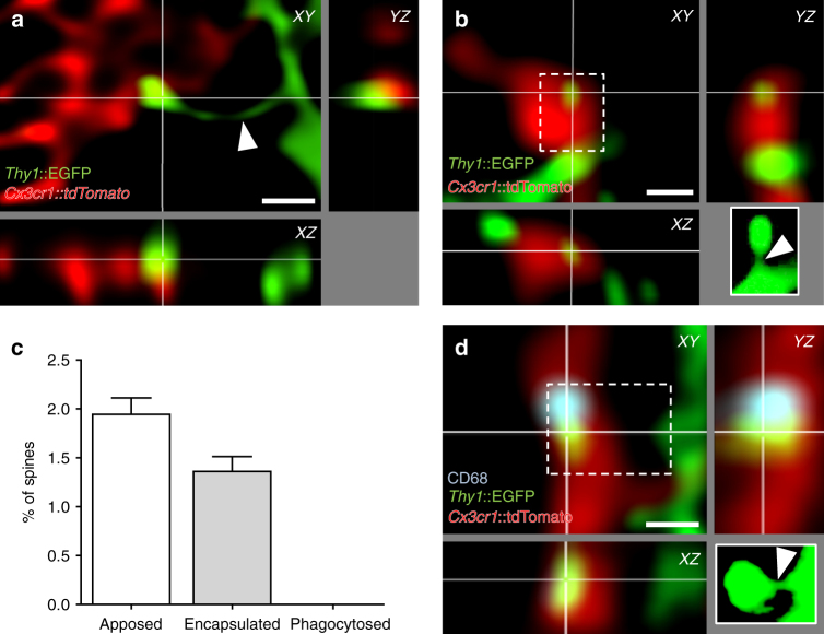
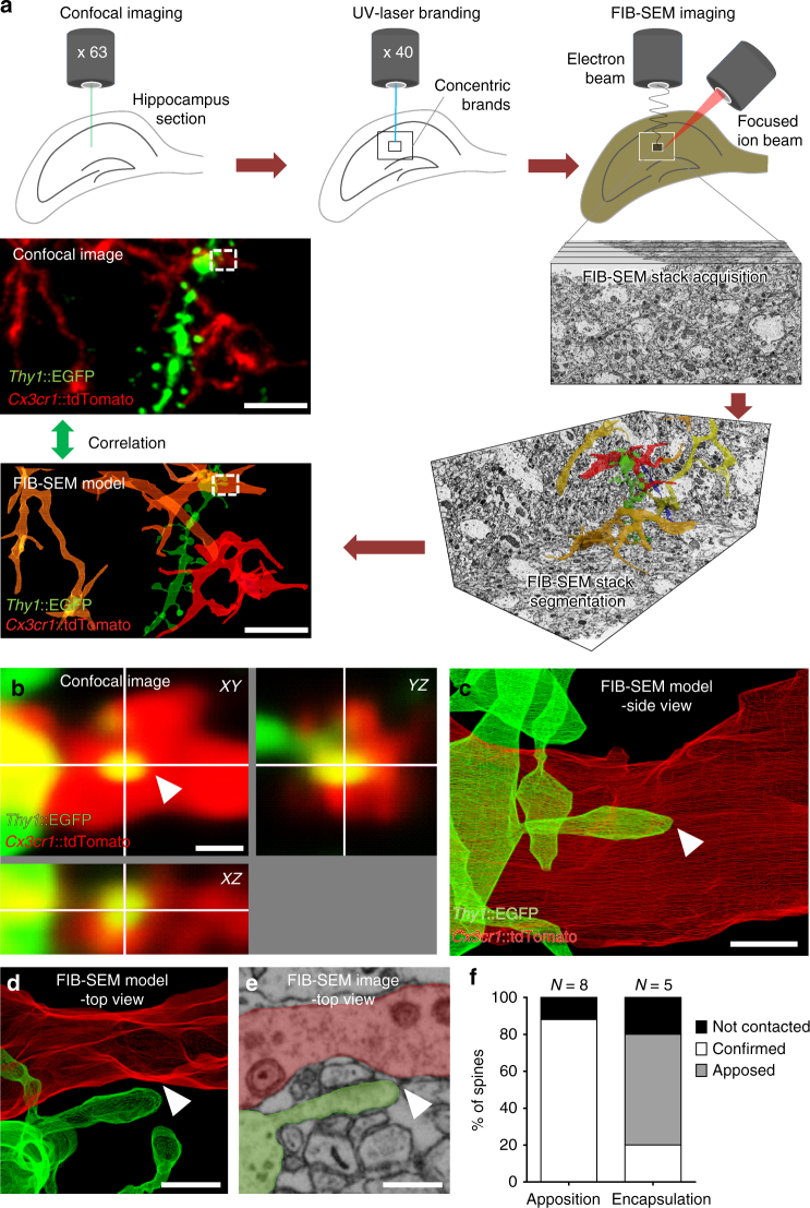
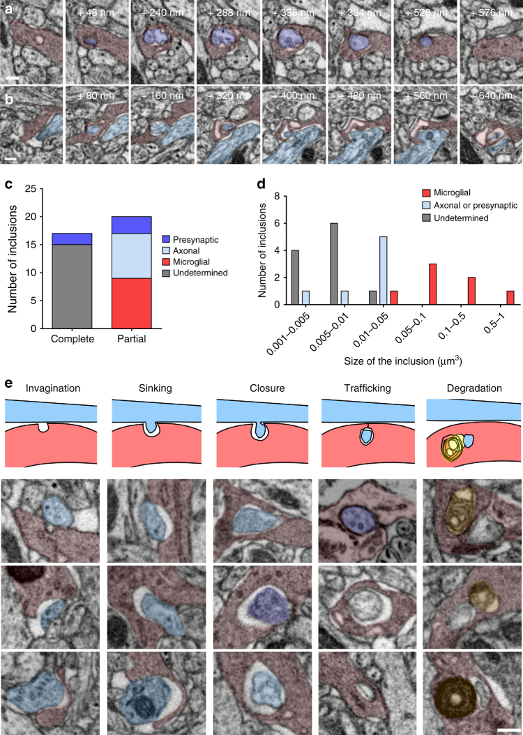
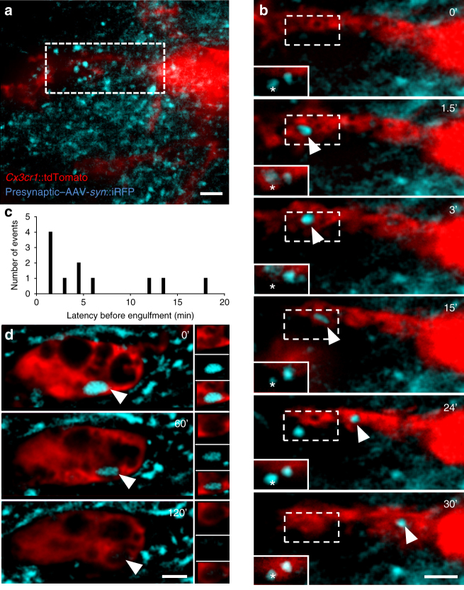
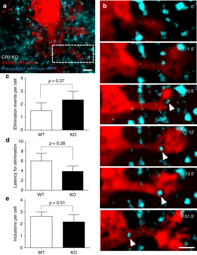
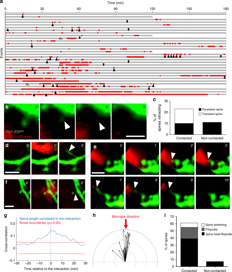
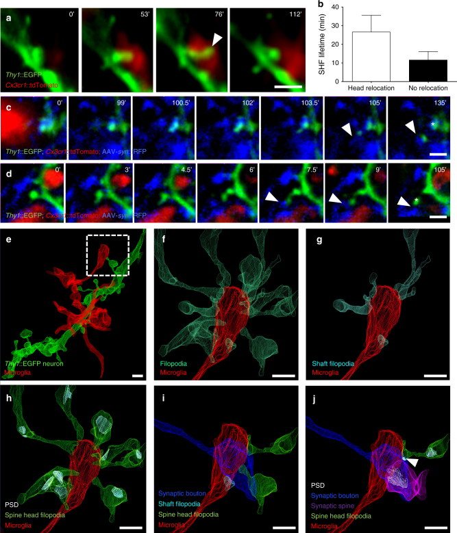
Microglia are highly motile glial cells that are proposed to mediate synaptic pruning during neuronal circuit formation. Disruption of signaling between microglia and neurons leads to an excess of immature synaptic connections, thought to be the result of impaired phagocytosis of synapses by microglia. However, until now the direct phagocytosis of synapses by microglia has not been reported and fundamental questions remain about the precise synaptic structures and phagocytic mechanisms involved. Here we used light sheet fluorescence microscopy to follow microglia-synapse interactions in developing organotypic hippocampal cultures, complemented by a 3D ultrastructural characterization using correlative light and electron microscopy (CLEM). Our findings define a set of dynamic microglia-synapse interactions, including the selective partial phagocytosis, or trogocytosis (trogo-: nibble), of presynaptic structures and the induction of postsynaptic spine head filopodia by microglia. These findings allow us to propose a mechanism for the facilitatory role of microglia in synaptic circuit remodeling and maturation.


Conflict of interest statement

The authors declare no competing interests.

Figures

Fig. 1
Fig. 1
Microglia do not phagocytose dendritic spines. Representative images of microglia (red, Cx3cr1::CreER; RC::LSL-tdTomato) a apposing or b encapsulating a dendritic spine (green, Thy1::EGFP; white insert: projection of the undeconvolved z-stack containing the contacted spine). Note that the neck of the contacted spine is intact (white arrow head). c Quantification of microglia–spine contacts. No spine was found phagocytosed, as contacted spines were always attached to their dendrite (n = 25 cells from 5 animals for 8944 spines analyzed, error bars are mean + SEM). d Encapsulated spine localizing next to a phagocytic compartment (blue, CD68 immunostaining). Scale bars: 0.5 µm
Fig. 2
Fig. 2
CLEM analysis of microglia-spine interactions. a Schematic of correlative light and electron microscopy (CLEM) workflow. b Confocal orthogonal view of a region of interest (ROI, dotted line in a) containing a spine encapsulated by microglia. c Segmentation of the ROI containing the encapsulation from the corresponding electron microscopy dataset, side view. d Top view of the ROI revealed that the spine was not encapsulated, with e no sign of elimination. f Quantification showed that the majority of the encapsulations observed by confocal microscopy were simple appositions by electron microscopy (n = 13 contacts analyzed from two animals). Scale bars: 5 µm for a, 0.5 µm for be
Fig. 3
Fig. 3
Microglia trogocytosis of presynaptic boutons and axons. Representative FIB-SEM image sequences of a complete presynaptic bouton inclusion (dark purple), as identified by its 40 nm vesicles content, inside microglia (red), b partial inclusion containing axonal material (clear blue) inside a microglia. c Quantification of microglial partial and complete inclusions (n = 37 inclusions from 8 cells, 4 animals). d Distribution of the volume of microglial inclusions. e Putative sequence of events leading to presynaptic bouton or axon material digestion by microglia, represented by a schematic and a collection of three examples for each step (gray: undetermined origin, yellow: lysosomes). Scale bars: 200 nm
Fig. 4
Fig. 4
Rapid trogocytosis of presynaptic material by microglia. a Low-magnification image of a stack projection of 35 consecutive optical sections (Δz = 0.48 µm) showing microglia (red, Cx3cr1::CreER; RC::LSL-tdTomato) surrounded by iRFP + presynaptic boutons from Schaffer collaterals (blue, AAV-Syn::iRFP) in the CA1 region of organotypic hippocampal cultures. b Time-lapse imaging revealed engulfment of a presynaptic bouton (single optical planes series from a, dotted box). The corresponding optical plane containing the presynaptic bouton (star) is shown in the plain white insert. Although most of the bouton has been internalized by the microglia and trafficked toward the soma (arrowhead), presynaptic material remains at the original site (star), indicating partial elimination. c Distribution of the latency to engulfment (n = 11 events from 8 cells originating from 3 organotypic slice cultures). d Representative image of an iRFP + inclusion in a microglia soma (arrowhead) showing slow degradation. Scale bars: 2 µm
Fig. 5
Fig. 5
CR3 is not necessary for microglia trogocytosis. a Low-magnification image of a stack-projection of 33 consecutive optical sections (Δz = 0.48 µm) showing a microglia (red, Cx3cr1::CreER; RC::LSL-tdTomato) surrounded by presynaptic boutons from Schaffer collaterals (blue, AAV-Syn::iRFP) in organotypic hippocampal slices from CR3 KO mice. b Time-lapse imaging of CR3 KO microglia–bouton interactions revealed engulfment of presynaptic material (single optical planes series from a, dotted box). No difference was found in the c number or d latency of microglia engulfment events, or e the number of iRFP inclusions per cell between WT and CR3 KO slices (two-sided unpaired t-test, n = 8 and 6 cells from 3 organotypic slice cultures, error bars are mean + SEM). Scale bars: 2 µm
Fig. 6
Fig. 6
Microglia induce spine head filopodia formation. a Quantification of microglia-spine contact duration (red) over the imaging session (gray). Each line represents a spine selected to have been contacted at least once by microglia and annotated for spine appearance (gray arrowhead), disappearance (black bar), and spine head filopodia formation (SHF, black arrowhead). Representative time sequence images of b spine disappearance, d filopodia formation, e SHF, and f spine stretching. c Quantification of spine retraction rate of contacted versus non-contacted neighboring spine. g Cross-correlation analysis revealed a significant increase in spine length during microglia-spine contact. h Vectorial analysis showed a significant correlation of microglial process direction (red arrow) with filopodia direction (Anderson–Darling test, P = 0.00047, n = 21 spine head filopodia analyzed, black arrows indicating filopodia length and direction, indentation = 1 µm). i Quantification of spine stretching, filopodia formation, and spine head filopodia formation events in contacted versus non-contacted neighboring spines (n = 31 and 28, respectively, from 4 organotypic slice cultures). Scale bars: 2 µm
Fig. 7
Fig. 7
Spine head filopodia-associated synapse remodeling. a Representative time sequence images of a spine head relocating to the tip of the SHF following its induction by microglia. b Quantification of SHF lifetime revealed more stable filopodia following relocation (n = 5 relocating SHF vs 16 non-relocating, analyzed from 3 organotypic slice cultures, error bars are mean + SEM). c,d SHF making a stable contact with a neighboring bouton (arrowhead). In c the spine persists at its original location (star), whereas in d the spine relocates to the newly contacted bouton (star). e Example of microglial process in contact with a spine extending a SHF (dotted box) as identified and visualized by CLEM. Further examination of the fully segmented EM dataset revealed f multiple filopodia extending toward the microglial process, of which g a few were simple filopodia and h the majority originated from mature spines bearing postsynaptic-densities (PSDs). It is noteworthy that the microglial process is in intimate contact with a presynaptic bouton and i several of the SHFs contact the same bouton, j one of which has formed an immature PSD (arrowhead, Supplementary Fig. 5) resulting in the formation of a multiple-synapse bouton. Scale bars: 1 µm

Comment in

  • Synaptic nibbling.
    Otto G. Otto G. Nat Rev Neurosci. 2018 Jun;19(6):322. doi: 10.1038/s41583-018-0008-1. Nat Rev Neurosci. 2018. PMID: 29717204 No abstract available.
  • Microglia: Picky Brain Eaters.
    Villani A, Peri F. Villani A, et al. Dev Cell. 2019 Jan 7;48(1):3-4. doi: 10.1016/j.devcel.2018.12.013. Dev Cell. 2019. PMID: 30620901

References

    1. Kettenmann H, Hanisch UK, Noda M, Verkhratsky A. Physiology of microglia. Physiol. Rev. 2011;91:461–553. doi: 10.1152/physrev.00011.2010. - DOI - PubMed
    1. Davalos D, et al. ATP mediates rapid microglial response to local brain injury in vivo. Nat. Neurosci. 2005;8:752–758. doi: 10.1038/nn1472. - DOI - PubMed
    1. Nimmerjahn A, Kirchhoff F, Helmchen F. Resting microglial cells are highly dynamic surveillants of brain parenchyma in vivo. Science. 2005;308:1314–1318. doi: 10.1126/science.1110647. - DOI - PubMed
    1. Dalmau I, Finsen B, Zimmer J, González B, Castellano B. Development of microglia in the postnatal rat hippocampus. Hippocampus. 1998;8:458–474. doi: 10.1002/(SICI)1098-1063(1998)8:5<458::AID-HIPO6>3.0.CO;2-N. - DOI - PubMed
    1. Mody M, et al. Genome-wide gene expression profiles of the developing mouse hippocampus. Proc. Natl Acad. Sci. USA. 2001;98:8862–8867. doi: 10.1073/pnas.141244998. - DOI - PMC - PubMed

Publication types

Substances