. 2018 Feb 8:2018:3786083.
doi: 10.1155/2018/3786083. eCollection 2018.

Study of the Influence of Age in 18F-FDG PET Images Using a Data-Driven Approach and Its Evaluation in Alzheimer's Disease

Affiliations

Study of the Influence of Age in 18F-FDG PET Images Using a Data-Driven Approach and Its Evaluation in Alzheimer's Disease

Jiehui Jiang et al. Contrast Media Mol Imaging. .

Abstract

Objectives: 18F-FDG PET scan is one of the most frequently used neural imaging scans. However, the influence of age has proven to be the greatest interfering factor for many clinical dementia diagnoses when analyzing 18F-FDG PET images, since radiologists encounter difficulties when deciding whether the abnormalities in specific regions correlate with normal aging, disease, or both. In the present paper, the authors aimed to define specific brain regions and determine an age-correction mathematical model.

Methods: A data-driven approach was used based on 255 healthy subjects.

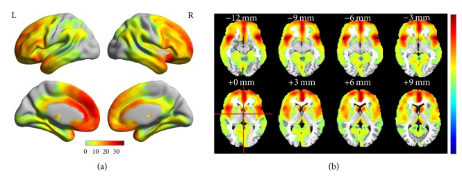
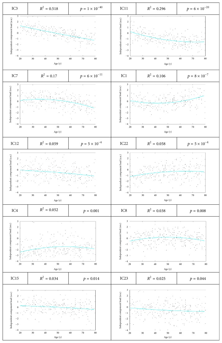
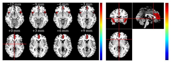
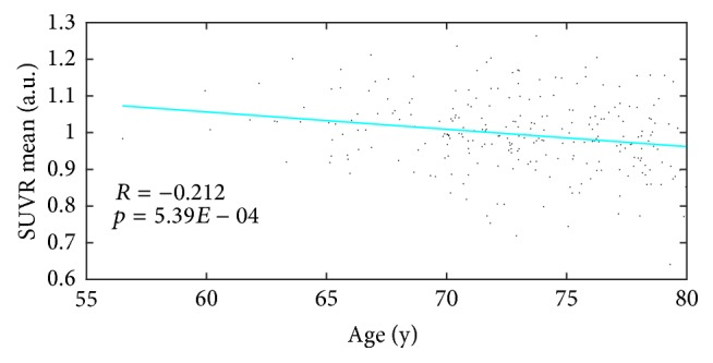
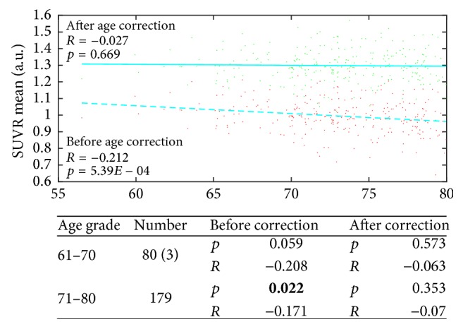
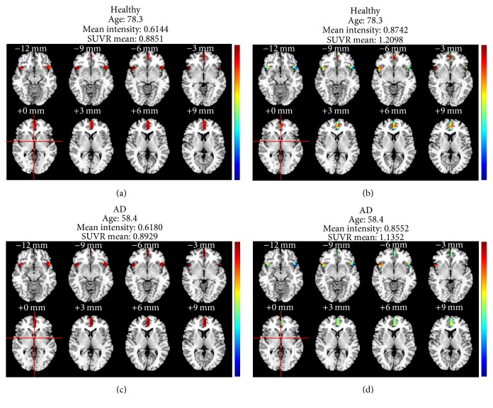
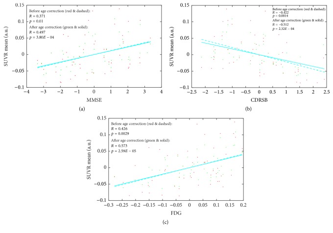
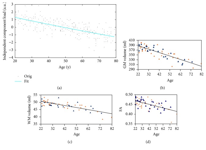
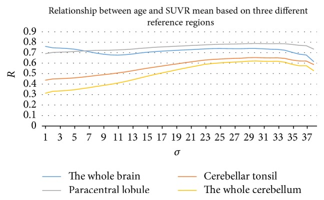
Results: The inferior frontal gyrus, the left medial part and the left medial orbital part of superior frontal gyrus, the right insula, the left anterior cingulate, the left median cingulate, and paracingulate gyri, and bilateral superior temporal gyri were found to have a strong negative correlation with age. For evaluation, an age-correction model was applied to 262 healthy subjects and 50 AD subjects selected from the ADNI database, and partial correlations between SUVR mean and three clinical results were carried out before and after age correction.

Conclusion: All correlation coefficients were significantly improved after the age correction. The proposed model was effective in the age correction of both healthy and AD subjects.


Figures

Figure 1
Figure 1
Anatomic structure of the brain regions corresponding to the significant age-related IC3 (pseudocolor) obtained across all 255 healthy subjects.
Figure 2
Figure 2
Plots of the post hoc relationship between the ICs and age.
Figure 3
Figure 3
Cumulative distribution of the voxel values.
Figure 4
Figure 4
Anatomic structure of the final brain region with σ = 30.
Figure 5
Figure 5
Scatter plot and fitting curves between SUVR mean formed from brain regions and age.
Figure 6
Figure 6
Scatter plot and fitting curves between SUVR mean and age across the 262 healthy subjects before (red and dashed) and after (green and solid) age correction. In addition, the statistical results of the age correction in the two age groups of the 252 healthy subjects are presented below.
Figure 7
Figure 7
Visual result demonstration of a qualitative case study.
Figure 8
Figure 8
Scatter plot and fitting curves between SUVR mean and (a) MMSE, (b) CDRSB, and (c) FDG across the 50 AD subjects before (red dots and dashed lines) and after (green dots and solid lines) age correction.
Figure 9
Figure 9
Similar linear scatter plots of negative correlations between age and different imaging indices. (a) Results from the present study. Correlation between SUVR mean and age. Correlations between (a) IC value (b; c; d) Results in [7]. Correlations between (b) GM volume, (c) WM volume, and (d) FA and age.
Figure 10
Figure 10
Relationship between age and SUVR mean based on four different reference regions: the cerebellum, the cerebellar tonsil, the paracentral lobule, and the whole brain region.

References

    1. Savva G. M., Wharton S. B., Ince P. G. Age, Neuropathology, and Dementia. The New England Journal of Medicine. 2009;360:2302–2309. doi: 10.1056/NEJMoa0806142. - DOI - PubMed
    1. Patterson B. W., Elbert D. L., Mawuenyega K. G., et al. Age and amyloid effects on human central nervous system amyloid-beta kinetics. Annals of Neurology. 2015;78(3):439–453. doi: 10.1002/ana.24454. - DOI - PMC - PubMed
    1. McKhann G., Drachman D., Folstein M., Katzman R., Price D., Stadlan E. M. Clinical diagnosis of Alzheimer's disease: report of the NINCDS-ADRDA work group * under the auspices of department of health and human services task force on alzheimer's disease. Neurology. 1984;34(7):939–944. doi: 10.1212/WNL.34.7.939. - DOI - PubMed
    1. Petrella J. R., Coleman R. E., Doraiswamy P. M. Neuroimaging and early diagnosis of alzheimer disease: A look to the future. Radiology. 2003;226(2):315–336. doi: 10.1148/radiol.2262011600. - DOI - PubMed
    1. Prince S. E., Woo S., Doraiswamy P. M., Petrella J. R. Functional MRI in the early diagnosis of Alzheimer's disease: is it time to refocus? Expert Review of Neurotherapeutics. 2008;8(2):169–175. doi: 10.1586/14737175.8.2.169. - DOI - PubMed

Publication types

Substances

LinkOut - more resources