

miR-143-3p regulates cell proliferation and apoptosis by targeting IGF1R and IGFBP5 and regulating the Ras/p38 MAPK signaling pathway in rheumatoid arthritis
- PMID: 29581736
- PMCID: PMC5863597
- DOI: 10.3892/etm.2018.5907
miR-143-3p regulates cell proliferation and apoptosis by targeting IGF1R and IGFBP5 and regulating the Ras/p38 MAPK signaling pathway in rheumatoid arthritis
Abstract
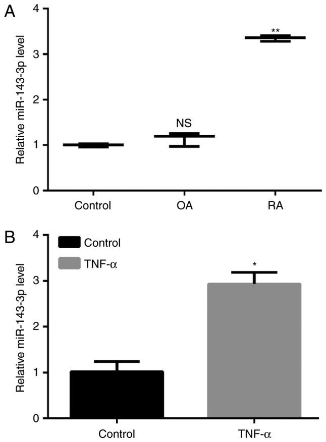
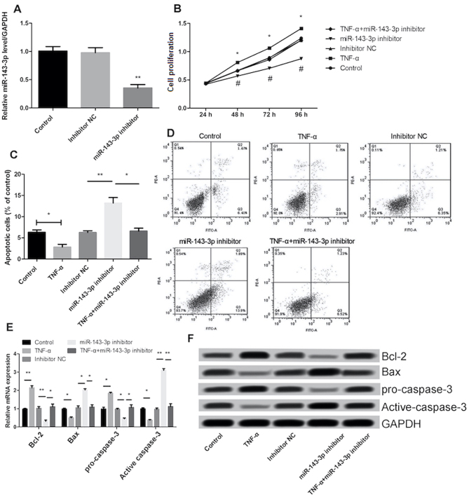
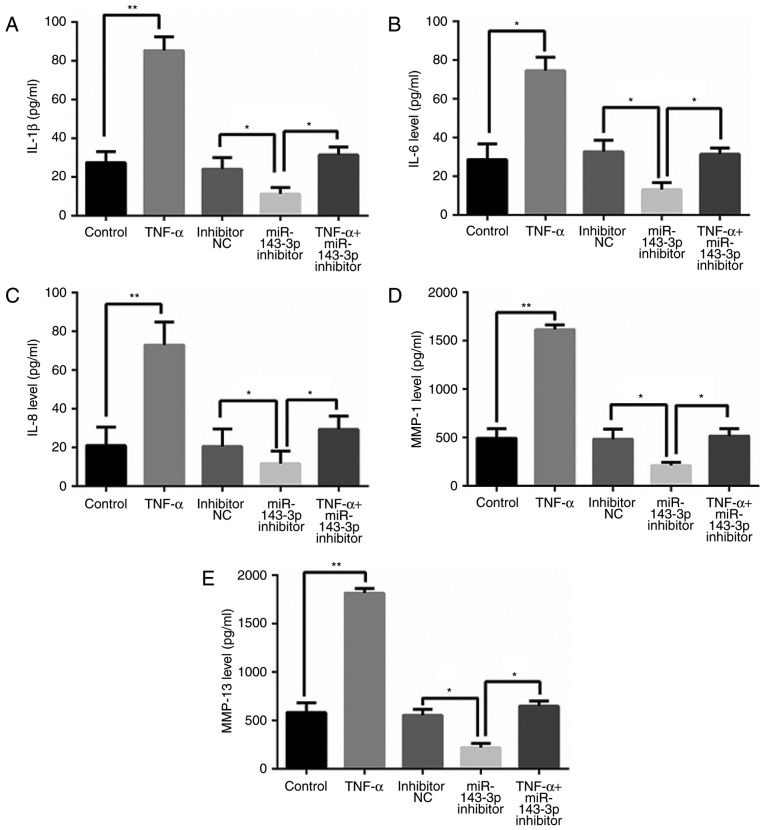
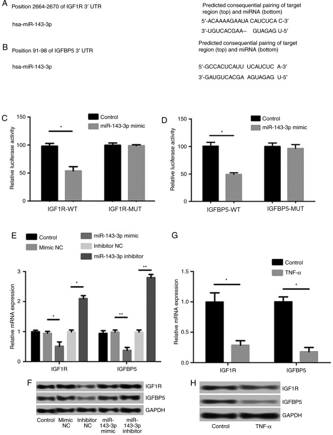
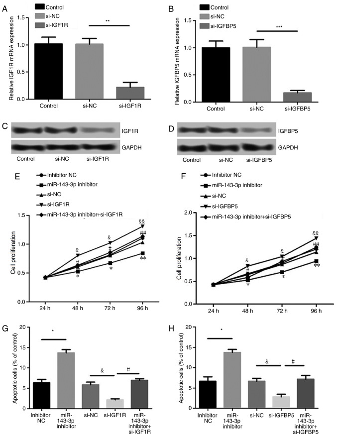
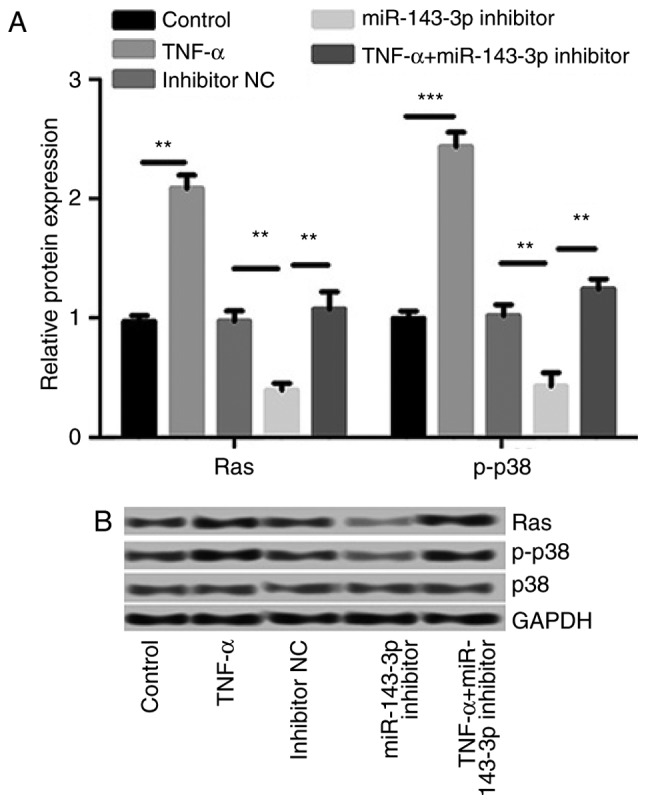
It has been demonstrated that the deregulation of microRNAs (miRNAs) affects the development of rheumatoid arthritis (RA). The primary objective of the current study was to determine the role of miR-143-3p in the progression of RA. The expression of miR-143-3p in synovium taken from patients with RA was assessed by reverse transcription-quantitative polymerase chain reaction. The expression of miR-143-3p was higher in synovium tissues of RA than that of osteoarthritis (OA). The decreased expression of miR-143-3p suppressed cell proliferation and promoted apoptosis in vitro. In addition, inhibition of miR-143-3p decreased levels of inflammatory cytokines, as determined by an enzyme-linked immunosorbent assay. IGF1R and IGFBP5 were found to be the target genes of miR-143-3p, and it was demonstrated that miR-143-3p regulated the proliferation and apoptosis of MH7A cells by targeting IGF1R and IGFBP5. Furthermore, TNF-α treatment stimulated the Ras/p38 mitogen activated protein kinase (MAPK) signaling pathway, whereas miR-143-3p inhibition suppressed it. The results of the current study indicate that miR-143-3p may regulate cell proliferation and apoptosis by targeting IGF1R and IGFBP5 expression and regulating the Ras/p38 MAPK signaling pathways. Therefore, miR-143-3p may be a novel therapeutic target in RA.
Keywords: insulin-like growth factor 1 receptor; insulin-like growth factor binding protein 5; microRNA-143-3p; rheumatoid arthritis; synovial fibroblasts.
Figures
Similar articles
-
MicroRNA-193a-3p participates in the progression of rheumatoid arthritis by regulating proliferation and apoptosis of MH7A cells through targeting IGFBP5.Eur Rev Med Pharmacol Sci. 2019 Jun;23(11):4850-4857. doi: 10.26355/eurrev_201906_18072. Eur Rev Med Pharmacol Sci. 2019. PMID: 31210318
-
miR-410-3p regulates proliferation and apoptosis of fibroblast-like synoviocytes by targeting YY1 in rheumatoid arthritis.Biomed Pharmacother. 2019 Nov;119:109426. doi: 10.1016/j.biopha.2019.109426. Epub 2019 Sep 7. Biomed Pharmacother. 2019. PMID: 31505424
-
MiR-199a-3p inhibits proliferation and induces apoptosis in rheumatoid arthritis fibroblast-like synoviocytes via suppressing retinoblastoma 1.Biosci Rep. 2018 Nov 13;38(6):BSR20180982. doi: 10.1042/BSR20180982. Print 2018 Dec 21. Biosci Rep. 2018. PMID: 30352835 Free PMC article.
-
MiR-223-3p in Cardiovascular Diseases: A Biomarker and Potential Therapeutic Target.Front Cardiovasc Med. 2021 Jan 20;7:610561. doi: 10.3389/fcvm.2020.610561. eCollection 2020. Front Cardiovasc Med. 2021. PMID: 33553260 Free PMC article. Review.
-
Research progress on the role and mechanism of miR-671 in bone metabolism and bone-related diseases.Front Oncol. 2023 Jan 11;12:1018308. doi: 10.3389/fonc.2022.1018308. eCollection 2022. Front Oncol. 2023. PMID: 36713572 Free PMC article. Review.
Cited by
-
The Role of Extracellular Vesicles in the Pathogenesis and Treatment of Rheumatoid Arthritis and Osteoarthritis.Cells. 2023 Nov 27;12(23):2716. doi: 10.3390/cells12232716. Cells. 2023. PMID: 38067147 Free PMC article. Review.
-
Low fluid shear stress promotes chondrocyte proliferation and extracellular matrix secretion by downregulating mir-143-3p and activating the ERK5/KLF4 signaling pathway.Sci Rep. 2024 Nov 12;14(1):27737. doi: 10.1038/s41598-024-78676-w. Sci Rep. 2024. PMID: 39532925 Free PMC article.
-
Rheumatoid arthritis treatment: Is exercise a game changer?Turk J Phys Med Rehabil. 2024 Nov 28;70(4):415-426. doi: 10.5606/tftrd.2024.16088. eCollection 2024 Dec. Turk J Phys Med Rehabil. 2024. PMID: 40028397 Free PMC article. Review.
-
Clinical effect and biological mechanism of exercise for rheumatoid arthritis: A mini review.Front Immunol. 2023 Jan 6;13:1089621. doi: 10.3389/fimmu.2022.1089621. eCollection 2022. Front Immunol. 2023. PMID: 36685485 Free PMC article. Review.
-
MiRNA Profiles of Extracellular Vesicles Secreted by Mesenchymal Stromal Cells-Can They Predict Potential Off-Target Effects?Biomolecules. 2020 Sep 22;10(9):1353. doi: 10.3390/biom10091353. Biomolecules. 2020. PMID: 32971982 Free PMC article.
References
-
- Okada Y, Naka K, Kawamura K, Matsumoto T, Nakanishi I, Fujimoto N, Sato H, Seiki M. Localization of matrix metalloproteinase 9 (92-kilodalton gelatinase/type IV collagenase=gelatinase B) in osteoclasts: Implications for bone resorption. Lab Invest. 1995;72:311–322. - PubMed
LinkOut - more resources
Full Text Sources
Other Literature Sources