

The Main Anatomical Variations of the Pancreatic Duct System: Review of the Literature and Its Importance in Surgical Practice
- PMID: 29581798
- PMCID: PMC5862083
- DOI: 10.14740/jocmr3344w
The Main Anatomical Variations of the Pancreatic Duct System: Review of the Literature and Its Importance in Surgical Practice
Abstract
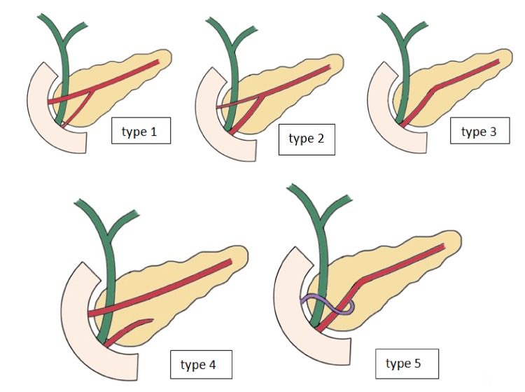
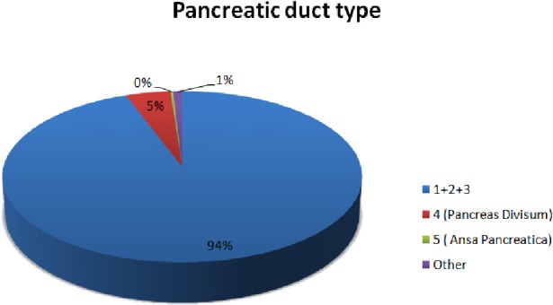
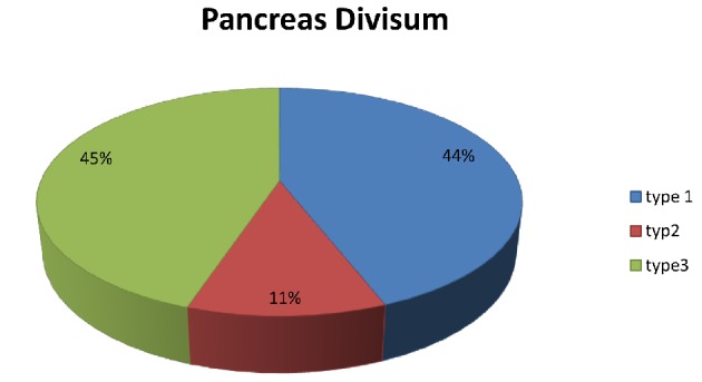
Anatomical variations or anomalies of the pancreatic ducts are important in the planning and performance of endoscopic retrograde cholangiopancreatography (ERCP) and surgical procedures of the pancreas. Normal pancreatic duct anatomy occurs in approximately 94.3% of cases, and multiple variations have been described for the remaining 5.7%. The purpose of this study was to review the literature on the pancreatic duct anatomy and to underline its importance in daily invasive endoscopic and surgical practice. Two main databases were searched for suitable articles published from 2000 to 2017, and results concerning more than 8,200 patients were included in the review. The most common anatomical variation was that of pancreas divisum, which appeared in approximately 4.5% of cases.
Keywords: Anatomic; Anatomy; Anomalies; Pancreatic duct; Variations.
Conflict of interest statement
The authors declare that they do not have any conflict of interest.
Figures
References
-
- Turkvatan A, Erden A, Turkoglu MA, Yener O. Congenital variants and anomalies of the pancreas and pancreatic duct: imaging by magnetic resonance cholangiopancreaticography and multidetector computed tomography. Korean J Radiol. 2013;14(6):905–913. doi: 10.3348/kjr.2013.14.6.905. - DOI - PMC - PubMed
-
- De Filippo M, Calabrese M, Quinto S, Rastelli A, Bertellini A, Martora R, Sverzellati N. et al. Congenital anomalies and variations of the bile and pancreatic ducts: magnetic resonance cholangiopancreatography findings, epidemiology and clinical significance. Radiol Med. 2008;113(6):841–859. doi: 10.1007/s11547-008-0298-x. - DOI - PubMed
Publication types
LinkOut - more resources
Full Text Sources
Other Literature Sources