. 2018 Jan 18:2018:1450828.
doi: 10.1155/2018/1450828. eCollection 2018.

Protein Kinase G Induces an Immune Response in Cows Exposed to Mycobacterium avium Subsp. paratuberculosis

Affiliations

Protein Kinase G Induces an Immune Response in Cows Exposed to Mycobacterium avium Subsp. paratuberculosis

Horacio Bach et al. Biomed Res Int. .

Abstract

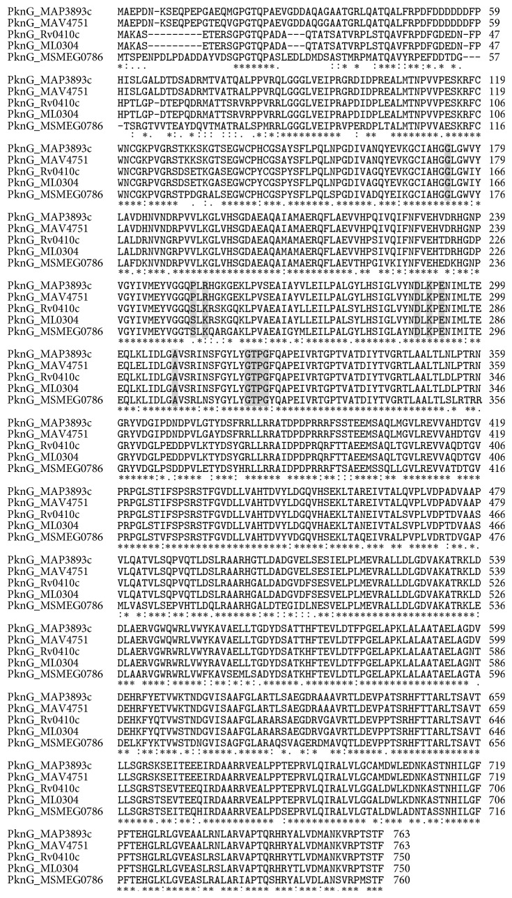
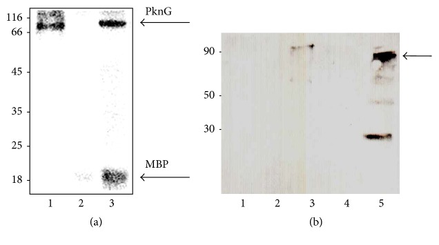
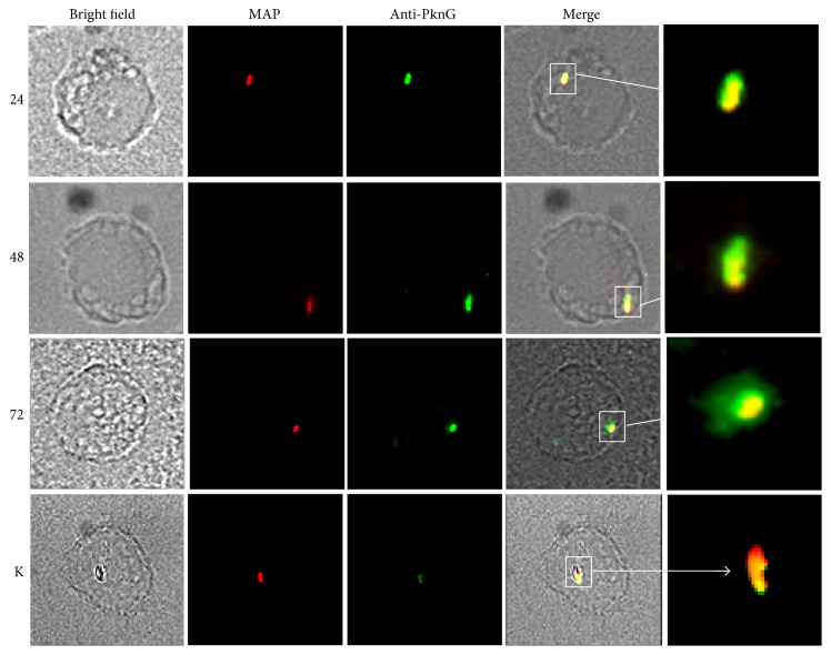
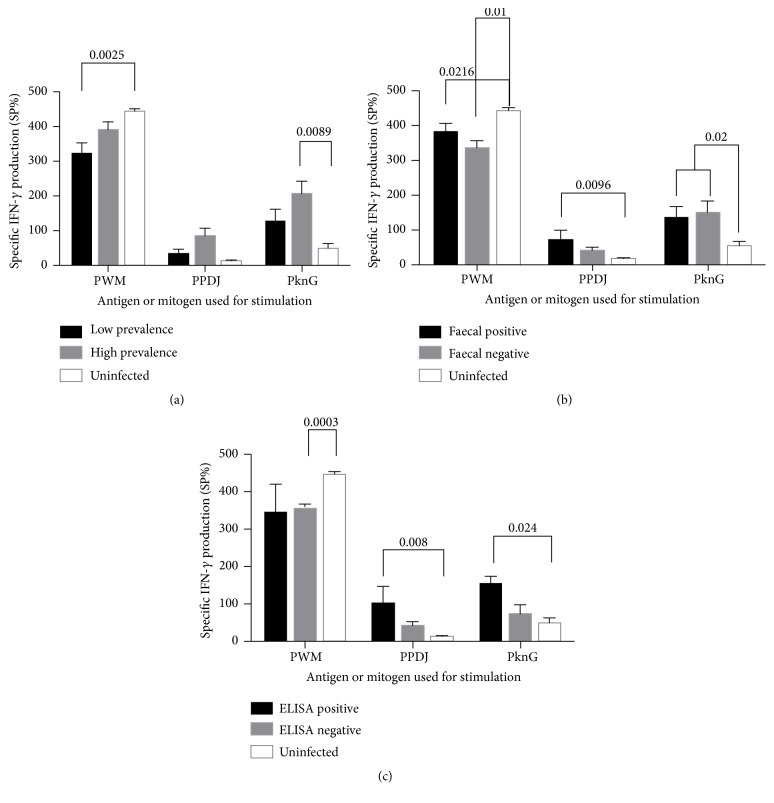
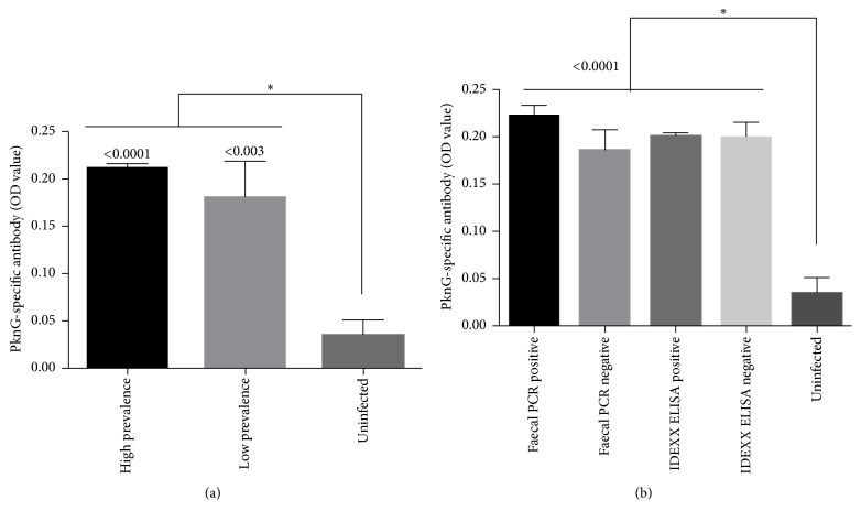
To establish infection, pathogens secrete virulence factors, such as protein kinases and phosphatases, to modulate the signal transduction pathways used by host cells to initiate immune response. The protein MAP3893c is annotated in the genome sequence of Mycobacterium avium subspecies paratuberculosis (MAP), the causative agent of Johne's disease, as the serine/threonine protein kinase G (PknG). In this work, we report that PknG is a functional kinase that is secreted within macrophages at early stages of infection. The antigen is able to induce an immune response from cattle exposed to MAP in the form of interferon gamma production after stimulation of whole blood with PknG. These findings suggest that PknG may contribute to the pathogenesis of MAP by phosphorylating macrophage signalling and/or adaptor molecules as observed with other pathogenic mycobacterial species.


Figures

Figure 1
Figure 1
MAP PknG alignment to mycobacterial PknGs. Selected mycobacterial PknG were aligned to MAP PknG. The PknG from the following Mycobacterium strains were aligned (locus tag, identity, similarity): M. avium subsp. paratuberculosis (MAP3893c), M. avium 104 (MAV4751, 86%. 92%), M. tuberculosis (Rv0410c, 86%, 91%), M. leprae (ML0304, 81%, 88%), and M. smegmatis (MSMEG0786, 79%, 81%). Letters in gray represent the substrate-binding residues.
Figure 2
Figure 2
Phosphorylation activity and immunoprecipitation of MAP PknG. (a) In vitro phosphorylation of MBP by MAP PknG. Recombinant his-tagged PknG was purified and incubated with [γ- 32P] ATP and MBP. Samples were separated by SDS-12% PAGE and stained with Coomassie blue followed by visualization by autoradiography. Lanes: 1, recombinant PknG; 2, MBP; 3, recombinant PknG + MBP. Molecular mass markers are indicated on the left in kDa. (b) The immunoprecipitants of infected and noninfected THP-1 cells were resolved by SDS-10% PAGE, electroblotted onto a nitrocellulose membrane, and exposed to rabbit anti-PknG antibodies. Lanes: 1, THP-1 cell-free lysate; 2, BSA (negative control); 3, recombinant PknG; 4, THP-1 cell lysate; 5, MAP-infected THP-1 lysate. Molecular mass markers in kDa; arrow points to the whole PknG.
Figure 3
Figure 3
PknG is secreted by MAP during infection. Fluorescence microscopy of infected THP-1 cells. THP-1 cells were infected with rhodamine 6G-labelled bacteria. Samples were obtained at 24, 48, and 72 h after infection and permeabilized with primary and secondary antibodies as described. PknG secretion was observed using fluorescence microscopy, where yellow indicates colocalization of the bacterium (in red) and the Alexa 488-anti-PknG antibodies (in green). Numbers on the left indicate hours. K, killed bacteria. White squares indicate phagosomes inside macrophages.
Figure 4
Figure 4
Measurement of IFN-γ secreted in whole blood exposed to mycobacterial antigens. Blood was stimulated with Pokeweed mitogen (PWM), PPDJ, or PknG. Cattle were categorized based on (a) prevalence of MAP infection on farms, (b) PCR diagnosis of faecal shedding of MAP, and (c) serotype according to IDEXX ELISA. Bars are representative of the mean ± SE.
Figure 5
Figure 5
Immunogenicity of PknG in cattle sera. (a) Sera from high and low prevalence or uninfected farms and (b) sera from cattle classified as positive or negative for faecal PCR and IDEXX ELISA. Shown is the mean ± SEM. p value < 0.05.

References

    1. Smith R. L., Grohn Y. T., Pradhan A. K., et al. A longitudinal study on the impact of Johne's disease status on milk production in individual cows. Journal of Dairy Science. 2009;92(6):2653–2661. doi: 10.3168/jds.2008-1832. - DOI - PubMed
    1. Johne’s disease on U.S. dairies, 1991-2007, (2008)
    1. Sweeney R. W. Transmission of paratuberculosis. Veterinary Clinics of North America: Food Animal Practice. 1996;12(2):305–312. doi: 10.1016/S0749-0720(15)30408-4. - DOI - PubMed
    1. Ellingson J. L. E., Anderson J. L., Koziczkowski J. J., et al. Detection of viable Mycobacterium avium subsp. paratuberculosis in retail pasteurized whole milk by two culture methods and PCR. Journal of Food Protection. 2005;68(5):966–972. doi: 10.4315/0362-028X-68.5.966. - DOI - PubMed
    1. Abubakar I., Myhill D., Aliyu S. H., Hunter P. R. Detection of Mycobacterium avium subspecies paratubercubsis from patients with Crohn's disease using nucleic acid-based techniques: a systematic review and meta-analysis. Inflammatory Bowel Diseases. 2008;14(3):401–410. doi: 10.1002/ibd.20276. - DOI - PubMed

MeSH terms

Substances

LinkOut - more resources