. 2018 Jan 18:2018:4071356.
doi: 10.1155/2018/4071356. eCollection 2018.

Computational Investigation on the Biomechanical Responses of the Osteocytes to the Compressive Stimulus: A Poroelastic Model

Affiliations

Computational Investigation on the Biomechanical Responses of the Osteocytes to the Compressive Stimulus: A Poroelastic Model

Liping Wang et al. Biomed Res Int. .

Abstract

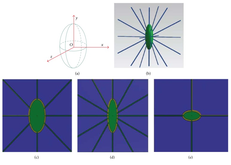
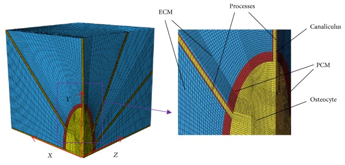
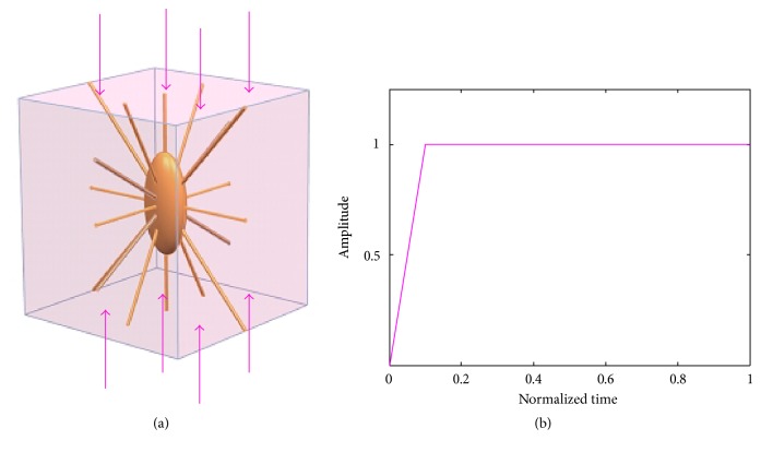
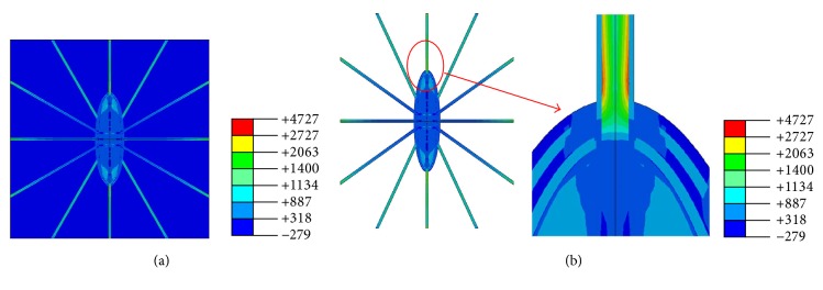
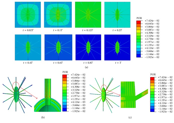
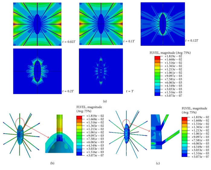
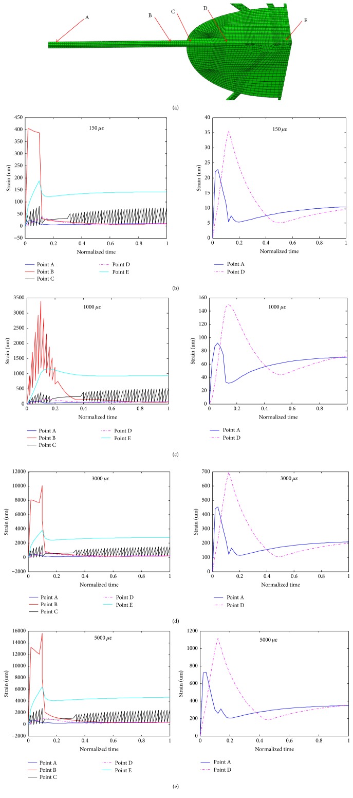
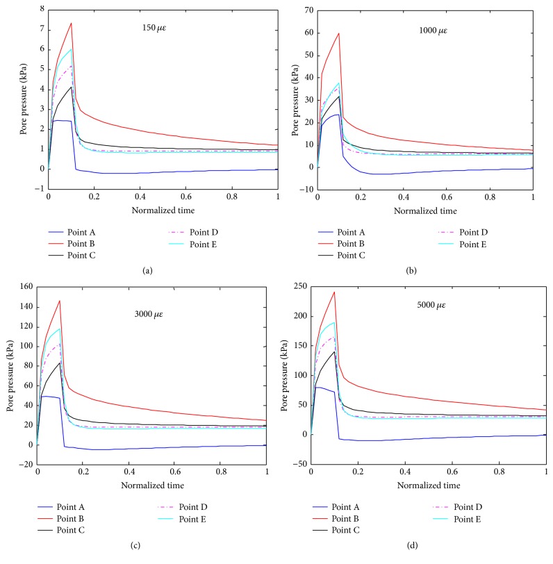
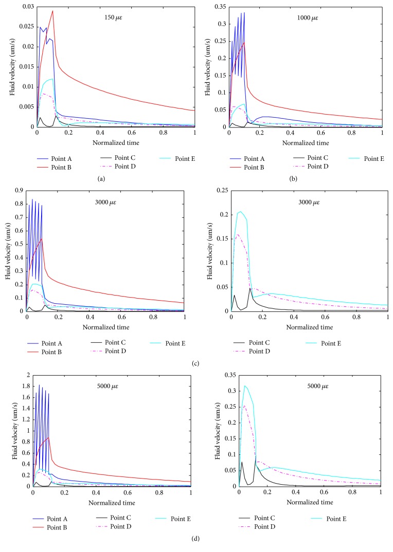
Osteocytes, the major type of bone cells embedded in the bone matrix and surrounded by the lacunar and canalicular system, can serve as biomechanosensors and biomechanotranducers of the bone. Theoretical analytical methods have been employed to investigate the biomechanical responses of osteocytes in vivo; the poroelastic properties have not been taken into consideration in the three-dimensional (3D) finite element model. In this study, a 3D poroelastic idealized finite element model was developed and was used to predict biomechanical behaviours (maximal principal strain, pore pressure, and fluid velocity) of the osteocyte-lacunar-canalicular system under 150-, 1000-, 3000-, and 5000-microstrain compressive loads, respectively, representing disuse, physiological, overuse, and pathological overload loading stimuli. The highest local strain, pore pressure, and fluid velocity were found to be highest at the proximal region of cell processes. These data suggest that the strain, pore pressure, and fluid velocity of the osteocyte-lacunar-canalicular system increase with the global loading and that the poroelastic material property affects the biomechanical responses to the compressive stimulus. This new model can be used to predict the mechanobiological behaviours of osteocytes under the four different compressive loadings and may provide an insight into the mechanisms of mechanosensation and mechanotransduction of the bone.


Figures

Figure 1
Figure 1
3-dimensional schematic representations of an idealized osteocyte-lacunar-canalicular system. (a) A schematic diagram of an osteocyte lacuna, where z is the intermediate axis, y the major axis, and x the minor axis of the lacuna in the local coordinate system. Schematic diagrams of osteocyte lacuna and canaliculi, showing (b) osteocyte cell body (green) with processes (blue); (c) in x-y plane; (d) in z-y plane; and (e) in x-z plane.
Figure 2
Figure 2
One-eighth symmetry model applied in the 3-dimensional finite element meshing analyses of the idealized osteocyte-lacunar-canalicular system, showing the geometric locations of extracellular matrix (ECM), perilacunar matrix (PCM), osteocyte cell body, canaliculi, and cell processes.
Figure 3
Figure 3
Conditions of compressive loading for the osteocyte-lacunar-canalicular system finite element model. (a) Uniaxial compressive loading; (b) loading mode.
Figure 4
Figure 4
Strain distributions of finite element model under 1000-microstrain global loading. (a) Perilacunar matrix and osteocyte; (b) extracellular matrix.
Figure 5
Figure 5
Pore pressure distributions of the osteocyte-lacunar-canalicular system under 1000-microstrain compressive loading. (a) The maximum pore pressure when t = 0.02, 0.1, 0.12, 0.2, 0.4, 0.6, 0.8, or 1.0T (total period of loading); (b) pore pressure distributions at canaliculi and perilacunar matrix when t = 0.1T; (c) pore pressure distributions in osteocyte and processes when t = 0.1T.
Figure 6
Figure 6
Fluid velocity distributions of the osteocyte-lacunar-canalicular system under the 1000-microstrain compressive loading. (a) Fluid velocities when t = 0.02, 0.1, 0.12, 0.2, and 1.0T, where T is the relative period of loading; (b) fluid velocity distribution at canaliculi and perilacunar matrix when t = 0.1T; and (c) fluid velocity distribution at the osteocyte and processes.
Figure 7
Figure 7
Detailed analyses of the maximum principal strains versus loading time at different locations (a) of the osteocyte cell body and a process when under different compressive loads ((b), (c), (d), and (e) for 150, 1000, 3000, and 5000 microstrains, respectively).
Figure 8
Figure 8
Detailed analyses of pore pressures versus loading time at different locations (defined in Figure 7(a)) of the osteocyte cell body and a process when the system was under different compressive loads ((a), (b), (c), and (d) for 150, 1000, 3000, and 5000 microstrains, respectively).
Figure 9
Figure 9
Detailed analyses of fluid velocities versus loading time at different locations (defined in Figure 7(a)) of the osteocyte cell body and a process when the system was under different compressive loads ((a), (b), (c), and (d) for 150, 1000, 3000, and 5000 microstrains, respectively).

References

    1. You L., Weinbaum S., Cowin S. C., Schaffler M. B. Ultrastructure of the osteocyte process and its pericellular matrix. The Anatomical Record. 2004;278A(2):505–513. doi: 10.1002/ar.a.20050. - DOI - PubMed
    1. Stern A. R., Nicolella D. P. Measurement and estimation of osteocyte mechanical strain. Bone. 2013;54(2):191–195. doi: 10.1016/j.bone.2013.01.037. - DOI - PubMed
    1. Rego E. B., Inubushi T., Kawazoe A., et al. Effect of PGE2 induced by compressive and tensile stresses on cementoblast differentiation in vitro. Archives of Oral Biolog. 2011;56(11):1238–1246. doi: 10.1016/j.archoralbio.2011.05.007. - DOI - PubMed
    1. Rath A. L., Bonewald L. F., Ling J., Jiang J. X., Van Dyke M. E., Nicolella D. P. Correlation of cell strain in single osteocytes with intracellular calcium, but not intracellular nitric oxide, in response to fluid flow. Journal of Biomechanics. 2010;43(8):1560–1564. doi: 10.1016/j.jbiomech.2010.01.030. - DOI - PMC - PubMed
    1. Bonewald L. F. Osteocytes as dynamic multifunctional cells. Annals of the New York Academy of Sciences. 2007;1116:281–290. doi: 10.1196/annals.1402.018. - DOI - PubMed

LinkOut - more resources