. 2018 Sep;59(3):363-374.
doi: 10.1165/rcmb.2017-0428OC.

Epithelial Heparan Sulfate Contributes to Alveolar Barrier Function and Is Shed during Lung Injury

Affiliations

Epithelial Heparan Sulfate Contributes to Alveolar Barrier Function and Is Shed during Lung Injury

Sarah M Haeger et al. Am J Respir Cell Mol Biol. 2018 Sep.

Abstract

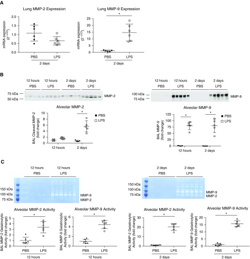
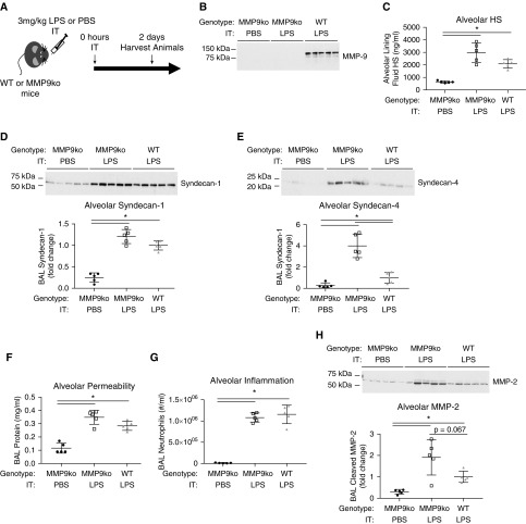
The lung epithelial glycocalyx is a carbohydrate-enriched layer lining the pulmonary epithelial surface. Although epithelial glycocalyx visualization has been reported, its composition and function remain unknown. Using immunofluorescence and mass spectrometry, we identified heparan sulfate (HS) and chondroitin sulfate within the lung epithelial glycocalyx. In vivo selective enzymatic degradation of epithelial HS, but not chondroitin sulfate, increased lung permeability. Using mass spectrometry and gel electrophoresis approaches to determine the fate of epithelial HS during lung injury, we detected shedding of 20 saccharide-long or greater HS into BAL fluid in intratracheal LPS-treated mice. Furthermore, airspace HS in clinical samples from patients with acute respiratory distress syndrome correlated with indices of alveolar permeability, reflecting the clinical relevance of these findings. The length of HS shed during intratracheal LPS-induced injury (≥20 saccharides) suggests cleavage of the proteoglycan anchoring HS to the epithelial surface, rather than cleavage of HS itself. We used pharmacologic and transgenic animal approaches to determine that matrix metalloproteinases partially mediate HS shedding during intratracheal LPS-induced lung injury. Although there was a trend toward decreased alveolar permeability after treatment with the matrix metalloproteinase inhibitor, doxycycline, this did not reach statistical significance. These studies suggest that epithelial HS contributes to the lung epithelial barrier and its degradation is sufficient to increase lung permeability. The partial reduction of HS shedding achieved with doxycycline is not sufficient to rescue epithelial barrier function during intratracheal LPS-induced lung injury; however, whether complete attenuation of HS shedding is sufficient to rescue epithelial barrier function remains unknown.

Keywords: acute respiratory distress syndrome; epithelial glycocalyx; heparan sulfate; syndecan.


Figures

Figure 1.
Figure 1.
Epithelial heparan sulfate (HS) degradation increases lung permeability but not inflammation. (A) Mice were instilled with 15 U intratracheal (IT) active or heat-inactivated (HI) heparinase I/III (Hep I/III or HI Hep I/III). (B) At 12 hours after instillation frozen lungs were sectioned and stained for HS (HS 10E4, green), and the peripheral airway/alveolar epithelium (Lycopersicon esculentum agglutinin [LEA] lectin, red). Scale bar: 100 μm. At 12, 24, and 72 hours after instillation, BAL fluid and plasma were obtained and (C and D) BAL and plasma HS concentration, (E) BAL protein, and (F) BAL albumin were measured. At 24 hours after instillation (G), lung wet/dry weight ratio was measured. At 12, 24, and 72 hours after instillation (H), BAL neutrophils were measured. (B) n = 3; (CH) n = 4–7. *P < 0.05.
Figure 2.
Figure 2.
Exogenous Hep I/III–generated HS fragments do not increase lung permeability or inflammation. (A) Exogenous HS (8.75 μg) was pretreated with 4.375 U of active or HI Hep I/III. The heparinase–HS mixture was then heat inactivated and intratracheally instilled. At 24 hours after intratracheal instillation of active or HI Hep I/III–treated HS, (B) BAL protein, (C) BAL neutrophils, and (D) whole lung TNF-α, IL-6, and IL-1β mRNA expression was measured. (BD) n = 4–5.
Figure 3.
Figure 3.
Increased airspace HS is detected during intratracheal LPS-induced lung injury in mice and in patients with acute respiratory distress syndrome (ARDS). (A) Mice were intratracheally instilled with 3 mg/kg LPS or PBS as control. At 12 hours and 2, 4, and 6 days after intratracheal instillation, (B) BAL protein was measured. (C) Hematoxylin and eosin staining was performed on lung sections from Day 2 after intratracheal LPS and PBS instillation. At 12 hours and 2, 4, and 6 days after intratracheal instillation, (D) BAL and (E) plasma HS was measured. Scale bars: 100 μm. (F) At 12 hours and 2 and 4 days after intratracheal instillation, BAL protein–HS correlation was analyzed by linear regression. (G) Protein and HS were measured in heat and moisture exchanger (HME) fluid from patients with ARDS, and HME protein–HS correlation was analyzed by linear regression. (B, D, and E) n = 3–9; (C) n = 3; (F) n = 10; (G) n = 15. *P < 0.05.
Figure 4.
Figure 4.
Increased airspace HS after intratracheal LPS instillation is long, and is accompanied by increased airspace syndecan-1 and syndecan-4. (A) Heparanase, reactive oxygen species (ROS), matrix metalloproteinase (MMP)-2, -7, and -9, and activation of disintegrin and metalloproteinase (ADAM)-17 cleave HS proteoglycans at distinct sites. (B) PAGE and Alcian blue/silver staining was performed on BAL fluid 2 and 4 days after intratracheal LPS instillation. dp = degree of polymerization; GAG = glycosaminoglycan. (C and D) BAL syndecan-1 and syndecan-4 were measured via Western blot from mice 2 days after intratracheal LPS instillation. (B) n = pool of 6–7 individual samples; (C and D) n = 5. *P < 0.05.
Figure 5.
Figure 5.
Lung MMP-9 mRNA expression and BAL MMP-2 and -9 protein and activity are increased after intratracheal LPS instillation. (A) Whole-lung MMP-2 and -9 mRNA expression was measured in mice 2 days after intratracheal LPS or PBS instillation. (B) BAL MMP-2 and -9 protein and (C) gelatinolytic activity were measured by Western blot and zymography in mice 12 hours and 2 days after intratracheal LPS or PBS instillation. (AC) n = 4–6. *P < 0.05.
Figure 6.
Figure 6.
Doxycycline partially inhibits the increase in airspace HS and syndecan-1, but not syndecan-4 or BAL protein, after intratracheal LPS instillation. (A) Mice were treated with 70 mg/kg oral doxycycline by gavage or water as control every 24 hours, starting 3 days before intratracheal instillation of LPS. Mice were harvested 2 days after intratracheal LPS instillation. BAL (B) HS, (C) syndecan-1, (D) syndecan-4, (E) protein, and (F) neutrophils were measured. (B) n = 9; (C and D) n = 6; (E) n = 9–10; (F) n = 10. *P < 0.05.
Figure 7.
Figure 7.
MMP-9 knockout (MMP9ko) mice are not protected from alveolar HS, syndecan-1, or syndecan-4 shedding, or BAL protein or neutrophilia, and exhibit increased BAL MMP-2 protein after intratracheal LPS instillation. (A) MMP9ko and wild-type (WT) mice were treated with intratracheal LPS, and MMP9ko mice were treated with intratracheal PBS as control. (B) A loss of BAL MMP-9 was confirmed by Western blot in MMP9ko mice in comparison to wild-type mice. BAL (C) HS, (D) syndecan-1, (E) syndecan-4, (F) protein, (G) neutrophils, and (H) MMP-2 protein were measured. (BH) n = 5. *P < 0.05.

References

    1. Lai M, Lampert IA, Lewis PD. The influence of fixation on staining of glycosaminoglycans in glial cells. Histochemistry. 1975;41:275–279. - PubMed
    1. Rambourg A, Neutra M, Leblond CP. Presence of a “cell coat” rich in carbohydrate at the surface of cells in the rat. Anat Rec. 1966;154:41–71. - PubMed
    1. Brooks RE. Ruthenium red stainable surface layer on lung alveolar cells; electron microscopic interpretation. Stain Technol. 1969;44:173–177. - PubMed
    1. Bignon J, Jaubert F, Jaurand MC. Plasma protein immunocytochemistry and polysaccharide cytochemistry at the surface of alveolar and endothelial cells in the rat lung. J Histochem Cytochem. 1976;24:1076–1084. - PubMed
    1. Dull RO, Dinavahi R, Schwartz L, Humphries DE, Berry D, Sasisekharan R, et al. Lung endothelial heparan sulfates mediate cationic peptide-induced barrier dysfunction: a new role for the glycocalyx. Am J Physiol Lung Cell Mol Physiol. 2003;285:L986–L995. - PubMed

Publication types

MeSH terms

LinkOut - more resources