. 2018 May 1;128(5):1772-1786.
doi: 10.1172/JCI97837. Epub 2018 Mar 26.

Role of proNGF/p75 signaling in bladder dysfunction after spinal cord injury

Affiliations

Role of proNGF/p75 signaling in bladder dysfunction after spinal cord injury

Jae Cheon Ryu et al. J Clin Invest. .

Abstract

Loss of bladder control is a challenging outcome facing patients with spinal cord injury (SCI). We report that systemic blocking of pro-nerve growth factor (proNGF) signaling through p75 with a CNS-penetrating small-molecule p75 inhibitor resulted in significant improvement in bladder function after SCI in rodents. The usual hyperreflexia was attenuated with normal bladder pressure, and automatic micturition was acquired weeks earlier than in the controls. The improvement was associated with increased excitatory input to the spinal cord, in particular onto the tyrosine hydroxylase-positive fibers in the dorsal commissure. The drug also had an effect on the bladder itself, as the urothelial hyperplasia and detrusor hypertrophy that accompany SCI were largely prevented. Urothelial cell loss that precedes hyperplasia was dependent on p75 in response to urinary proNGF that is detected after SCI in rodents and humans. Surprisingly, death of urothelial cells and the ensuing hyperplastic response were beneficial to functional recovery. Deleting p75 from the urothelium prevented urothelial death, but resulted in reduction in overall voiding efficiency after SCI. These results unveil a dual role of proNGF/p75 signaling in bladder function under pathological conditions with a CNS effect overriding the peripheral one.

Keywords: Neuroscience; Urology.


Conflict of interest statement

Conflict of interest: The authors have declared that no conflict of interest exists.

Figures

Figure 1
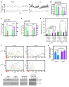
Figure 1. ProNGF is released into the urine in rodents and humans within hours of SCI.
(A) ProNGF is released into the urine almost immediately after spinal cord transection in mice. The blots were probed with anti-proNGF and pan-NGF antibodies. There was little mature NGF detectable in the urine at these time points. (B) Quantification of proNGF by Western blotting. Within-subject comparison was significant at P = 0.003 based on repeated measures of 1-way ANOVA after Greenhouse-Geisser correction, which resulted in P < 0.01. Subsequent pairwise comparisons were made using Bonferroni correction. (C) Quantification of proNGF detected in mouse urine by proNGF-specific ELISA. Note that mature NGF was not detected by mature NGF-specific ELISA. (D) Immunoprecipitation/Western analyses of urine samples illustrate that the bands detected with proNGF antibody in A are indeed proNGF. Mouse urine samples were immunoprecipitated with 27/21 mature NGF antibody and probed with proNGF antibody. (E) ProNGF is detected in the urine after contusion injuries in mice. (F) ProNGF is present in the urine after spinal cord transection in rats. Mature NGF was not detected by mature NGF-specific ELISA. (G) ProNGF was also detected in human urine after SCI. The upper blot was probed with proNGF-specific antibody (*artifact), while the lower blot was probed with pan-NGF antibody (H-20). Note that recombinant proNGF and mature NGF were included as controls. Based on human proNGF-specific ELISA, the amount of proNGF was 1 and 46 pg/mg for 3 and 6 hours after injury, respectively. Note that 3- and 6-hour urine samples were collected from 2 different individuals. ELISA assays also failed to detect mature NGF as in Western blotting. C, urine from healthy control without SCI.
Figure 2
Figure 2. p75 is responsible for umbrella cell apoptosis in response to urinary proNGF after SCI.
(A) Uroplakin-positive (UP+) umbrella cells are the major cell types that undergo apoptosis at 7 hours after SCI. The bladders were processed for TUNEL reaction and subsequently stained with UP or p75 antibodies. Urothelial and muscle layers of the bladder wall are indicated by U and M, respectively, and the bladder lumen is indicated by L. Scale bar: 150 μm. (B) p75 is responsible for umbrella cell apoptosis after SCI as indicated by the lack of TUNEL+ cells in the p75KO. The data were analyzed using repeated measures of 2-way ANOVA after Greenhouse-Geisser correction, which resulted in P < 0.001 comparing the vehicle and LM11A-31 treatments and the WT and p75-null mice. Subsequent pairwise comparisons were made using Bonferroni correction. (C) Intravesical instillation of proNGF blocking antibody blocked umbrella cell apoptosis completely at 7 hours after injury. The data were analyzed by Student’s t tests.
Figure 3
Figure 3. Hexagonal morphology of umbrella cells is preserved in p75KO mice and with LM11A-31 administration after SCI.
(A) Without injury, the luminal surface of the bladder exhibited the well-characterized polyhedral morphology with ridges that demarcate cell boundaries (blue lines and arrows; ref. 15). At 7 hours after injury, however, the surface morphology was drastically altered, with loss of the characteristic junctional ridges. By 2 days postinjury (dpi), umbrella cells were regenerated, but they were smaller in size than those without injury, although they established polyhedral morphology (red arrows). (B) With intravesical instillation of LM11A-31 immediately after SCI, however, umbrella cell loss was completely blocked, with intact polyhedral ridges at both 7 hours and 2 days after injury (blue lines and arrows). (C) At 7 hours after injury, the luminal surface of the bladder from p75KO mice was visually indistinguishable from that of the control bladder. Blue dotted lines mark the hexagonal boundaries of umbrella cells. Higher-magnification images illustrate the cell boundaries between neighboring cells (arrows). (D) H&E staining of the control and LM11A-31–treated bladders. Note the preservation of the urothelium at 1 day after injury with LM11A-31, while very few urothelial cells are visible in vehicle-treated bladder. LM11A-31 also prevented chronic urothelial hyperplasia and detrusor hypertrophy of the bladder at 10 days after injury. The bladder morphology appears similar to that of the uninjured control bladder. Urothelial and muscle layers of the bladder wall are indicated by U and M, respectively, and the bladder lumen is indicated by L. Scale bar: 250 μm. Experiments were performed 3 times, and representative ones are shown.
Figure 4
Figure 4. Selective deletion of p75 among umbrella cells results in complete block of umbrella cell apoptosis.
(A) Diagram of the targeting vectors in p75Δ-UP3a and p75c-UP3a mice. Red triangles represent loxP sites, and green P1 and P2 arrows represent PCR primers used in B. (B) Selective deletion of p75 in urothelial cells. Urothelial cells from tamoxifen-treated p75Δ-UP3a and p75c-UP3a mice were scraped into a tube, and the isolated genomic DNA was subjected to PCR using P1 and P2 primers. The primers generate PCR product only when Cre is activated. Note that the PCR band is present only in umbrella cells and not in the bladder minus urothelium in p75Δ-UP3a mice, indicating a selective p75 deletion in the urothelium. (C) p75 is not detected in umbrella cells (U), while it is clearly present in the muscle (M). L, bladder lumen. Scale bar: 150 μm. (D) Umbrella cell apoptosis was completely blocked in p75Δ-UP3a compared with that in p75c-UP3a mice.
Figure 5
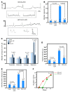
Figure 5. Selective deletion of p75 among umbrella cells influences micturition.
(A) Representative recordings of intravesical pressure during continuous intravesical infusion of saline and an open urethral outlet in conscious, unrestrained p75c-UP3a and p75Δ-UP3a mice with no SCI and SCI (2 weeks). (B) Overall voiding efficiency in no-SCI p75Δ-UP3a and p75c-UP3a mice was similar, although after SCI, p75Δ-UP3a mice exhibited worse bladder function than p75c-UP3a mice with a 32% drop in voiding efficiency (n = 3–5). (C and D) Under no-SCI condition, p75Δ-UP3a mice had significantly decreased infused volumes (IVs) that induced micturition and intermicturition intervals compared with those observed in p75c-UP3a mice. SCI significantly reduced intermicturition intervals (C) and IVs (D) in p75c-UP3a mice, and these effects were reversed in p75Δ-UP3a mice. Note that left y axes represent no SCI, while right y axes represent SCI groups. (E) No-SCI p75Δ-UP3a mice exhibited significantly increased average, minimum, and threshold bladder pressures compared with control, with no change in maximum micturition pressure. Comparisons among groups were made using ANOVA. When F ratios exceeded the adjusted critical value (P ≤ 0.0125), Bonferroni’s multiple-comparisons test was used to compare means among groups. (F) ProNGF/p75 signaling negatively influences TrpV4-mediated Ca2+ flux in primary mouse urothelial cells. Urothelial cells were incubated with 10 ng/ml of proNGF or vehicle for 30 minutes before Fluo-4 AM loading and GSK1016790A addition at 50 nM (red arrows; n = 4 independent experiments). (G) Quantification of the average peak amplitude of ΔF/F. Comparisons among groups were made using ANOVA, and pairwise comparisons were made by Tukey’s multiple-comparisons test (3–8 cells were included per treatment). (H) GSK1016790A facilitates cell-surface targeting of TrpV4 in HEK293T cells, which is inhibited by proNGF. ProNGF-mediated inhibition is lifted with LM11A-31 preincubation.
Figure 6
Figure 6. Systemic LM11A-31 greatly improves bladder function after SCI in mice.
(A) Representative bladder function recordings in conscious, unrestrained vehicle- or LM11A-31–treated mice with continuous intravesical instillation of saline with an open outlet at 28 days after injury. Upper recordings were from SCI mice that were treated with vehicle, while lower ones were from SCI mice that were treated with 100 mg/kg LM11A-31. Boxed areas: Representative single-fill cystometry recordings (arrow, infusion start) in conscious, unrestrained vehicle- or LM11A-31–treated mice from 2 different experiments. (B) After SCI, LM11A-31 significantly reduced bladder capacity, reaching that of spinal-intact mice. (C) Micturition pressures (threshold, average, maximum) were significantly reduced compared with those in vehicle-treated mice. (D and E) Infused volume and intermicturition intervals were reduced compared with those in vehicle-treated mice in both spinal-intact and SCI groups. (F) Greater numbers of mice treated with LM11A-31 recovered automatic micturition significantly earlier than vehicle-treated mice after SCI. Time (days) to recovery of automatic micturition for 2 groups was compared using a 2-tailed unpaired Student’s t test. P ≤ 0.05 was considered statistically significant. Comparisons of cystometric parameters among groups (BE) were made using ANOVA. When F ratios exceeded the adjusted critical value (P ≤ 0.0125), Bonferroni’s multiple-comparisons test was used to compare means among groups.
Figure 7
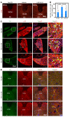
Figure 7. Colocalization of tyrosine hydroxylase among p75+ neurons in DRG and L6/S1 spinal cords after SCI.
(A) LM11A-31 reduced the number of c-fos+ cells in L6 spinal cords after SCI. DCM, dorsal commissure; cc, central canal; MDH, medial dorsal horn. Scale bar: 150 μm. (B) Quantification of A. Comparisons among groups were made using 1-way ANOVA (P ≤ 0.001), and pairwise comparisons were made by Tukey’s multiple-comparisons test. (C) p75 is expressed among tyrosine hydroxylase–positive (TH+) sensory neurons in L6/S1 DRG. DRGs were processed for double immunohistochemistry for TH and p75. Scale bars: 150 μm. The boxed areas are shown in the far right column; scale bars: 37.5 μm. (D) Colocalization of TH and p75 in fibers and neuronal soma in the DCM of L6/S1 spinal cords. Scale bars: 150 μm. The boxed areas are shown in the far right column; scale bars: 37.5 μm. Experiments were performed 3 times, and representative ones are shown.
Figure 8
Figure 8. Systemic LM11A-31 increases glutamatergic inputs after SCI in mice.
(A) Representative images of TH and vGlut1 immunostaining of L6/S1 spinal cords. Scale bars: 75 μm. The boxed areas are shown in 3 columns to the right; scale bars: 30 μm. (B) LM11A-31 increased the vGlut1+ synaptic puncta. Comparisons among groups were made using 1-way ANOVA, and pairwise comparisons were made by Tukey’s multiple-comparisons test. (C) LM11A-31 increased glutamatergic inputs to TH+ fibers. Comparisons among groups were made using 1-way ANOVA (P ≤ 0.05), and pairwise comparisons were made by Tukey’s multiple-comparisons test.

References

    1. Fowler CJ, Griffiths D, de Groat WC. The neural control of micturition. Nat Rev Neurosci. 2008;9(6):453–466. doi: 10.1038/nrn2401. - DOI - PMC - PubMed
    1. de Groat WC, Yoshimura N. Mechanisms underlying the recovery of lower urinary tract function following spinal cord injury. Prog Brain Res. 2006;152:59–84. doi: 10.1016/S0079-6123(05)52005-3. - DOI - PubMed
    1. Ochodnicky P, Cruz CD, Yoshimura N, Cruz F. Neurotrophins as regulators of urinary bladder function. Nat Rev Urol. 2012;9(11):628–637. doi: 10.1038/nrurol.2012.178. - DOI - PubMed
    1. Ochodnický P, Cruz CD, Yoshimura N, Michel MC. Nerve growth factor in bladder dysfunction: contributing factor, biomarker, and therapeutic target. Neurourol Urodyn. 2011;30(7):1227–1241. - PubMed
    1. Seki S, et al. Suppression of detrusor-sphincter dyssynergia by immunoneutralization of nerve growth factor in lumbosacral spinal cord in spinal cord injured rats. J Urol. 2004;171(1):478–482. doi: 10.1097/01.ju.0000088340.26588.74. - DOI - PubMed

Publication types

MeSH terms