. 2018 May 1;128(5):1807-1819.
doi: 10.1172/JCI99088. Epub 2018 Mar 26.

PAI1 mediates fibroblast-mast cell interactions in skin fibrosis

Affiliations

PAI1 mediates fibroblast-mast cell interactions in skin fibrosis

Neha Pincha et al. J Clin Invest. .

Abstract

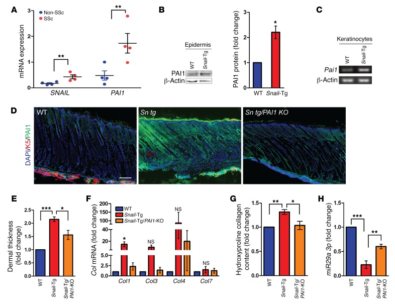
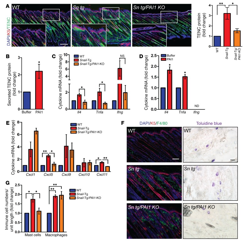
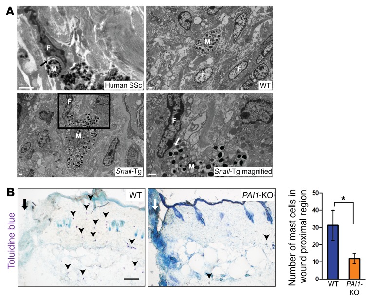
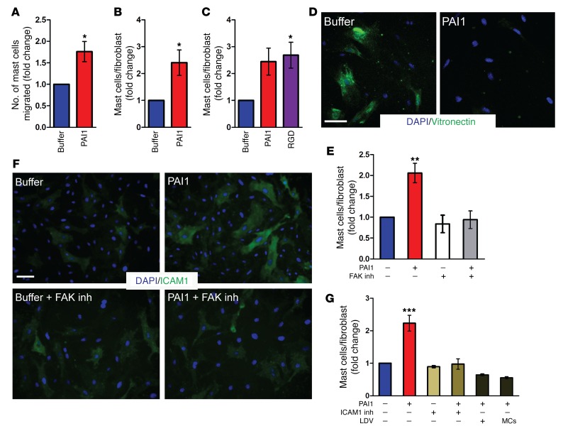
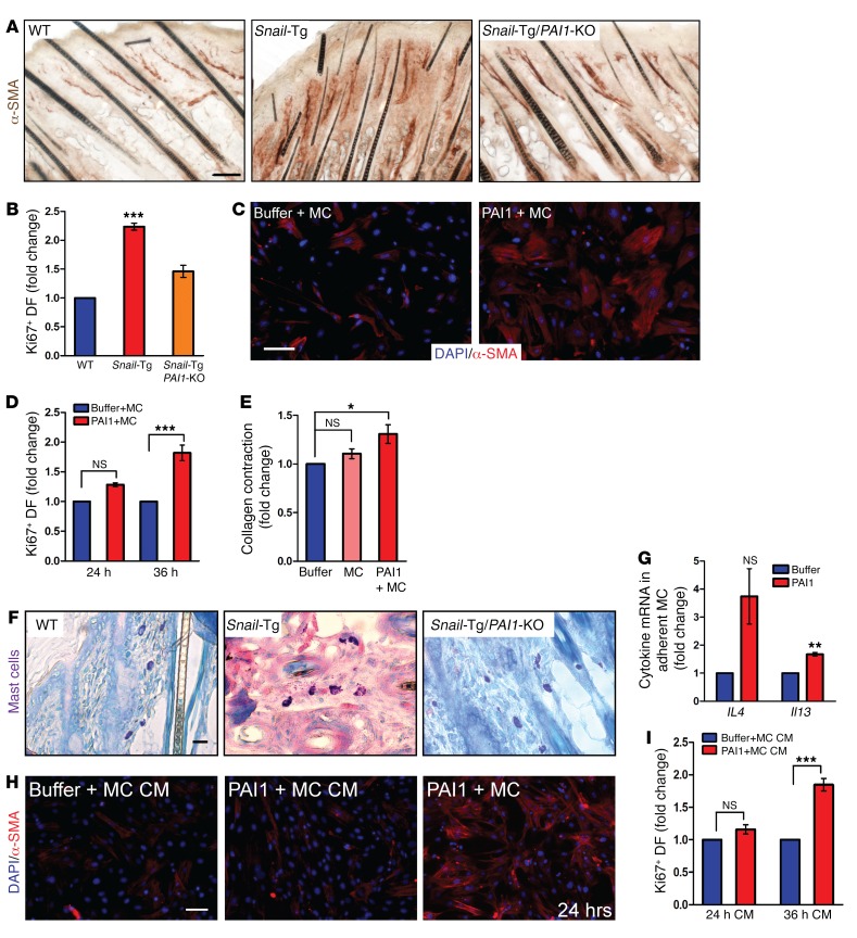
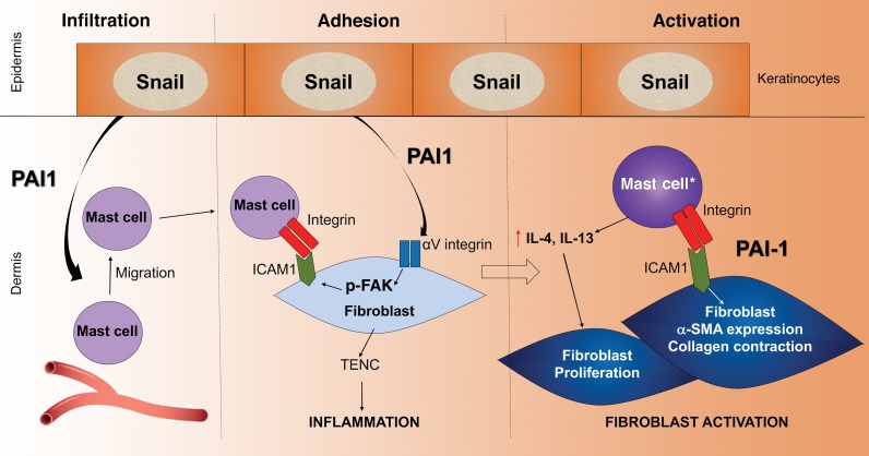
Fibrosis is a prevalent pathological condition arising from the chronic activation of fibroblasts. This activation results from the extensive intercellular crosstalk mediated by both soluble factors and direct cell-cell connections. Prominent among these are the interactions of fibroblasts with immune cells, in which the fibroblast-mast cell connection, although acknowledged, is relatively unexplored. We have used a Tg mouse model of skin fibrosis, based on expression of the transcription factor Snail in the epidermis, to probe the mechanisms regulating mast cell activity and the contribution of these cells to this pathology. We have discovered that Snail-expressing keratinocytes secrete plasminogen activator inhibitor type 1 (PAI1), which functions as a chemotactic factor to increase mast cell infiltration into the skin. Moreover, we have determined that PAI1 upregulates intercellular adhesion molecule type 1 (ICAM1) expression on dermal fibroblasts, rendering them competent to bind to mast cells. This heterotypic cell-cell adhesion, also observed in the skin fibrotic disorder scleroderma, culminates in the reciprocal activation of both mast cells and fibroblasts, leading to the cascade of events that promote fibrogenesis. Thus, we have identified roles for PAI1 in the multifactorial program of fibrogenesis that expand its functional repertoire beyond its canonical role in plasmin-dependent processes.

Keywords: Cell Biology; Fibrosis; Inflammation; Innate immunity; Skin.


Conflict of interest statement

Conflict of interest: The authors have declared that no conflict of interest exists.

Figures

Figure 1
Figure 1. PAI1 contributes to fibrosis in Snail-Tg skin.
(A) qPCR for SNAIL and PAI1 in skin samples from healthy individuals (Non-SSc) and scleroderma patients (SSc) (n = 4). (B) Western blot for PAI1 in WT and Snail-Tg epidermis and quantification. β-Actin was used as a loading control (n = 3). (C) Reverse transcriptase PCR of Pai1 in WT and Snail-Tg keratinocytes (n = 3). (D) WT, Snail-Tg (Sn tg), and Snail-Tg/Pai1-KO (Sn tg/PAI1 KO) neonatal skin analyzed for PAI1 secretion by immunostaining (n = 3). Scale bar: 50 μm. (EH) WT, Snail-Tg, and Snail-Tg/Pai1-KO adult skin sections were analyzed for (E) dermal thickness by quantitation of H&E-stained sections (n = 4); (F) Col1, -3, -4, and -7 by qPCR (n = 3); (G) collagen protein levels by hydroxyproline assay (n = 5); and (H) miR29a 3p levels by qPCR (n = 3). Data represent the mean ± SEM. *P < 0.05, **P < 0.01, and ***P < 0.001, by Student’s t test (A and B) and 1-way ANOVA followed by Tukey’s post hoc analysis (EH).
Figure 2
Figure 2. PAI1 affects fibrogenesis during the inflammation phase in Snail-Tg skin.
(A) WT, Snail-Tg, and Snail-Tg/Pai1-KO pup skin sections were analyzed for TENC localization by immunostaining (n = 3; scale bar and original magnification: 50 μm) and total TENC protein levels by Western blotting (n = 3). (B) Quantification of secreted TENC in recombinant PAI1-treated fibroblast conditioned media (n = 3). qPCR for proinflammatory cytokines Tnfa, Il4, and Ifng in (C) neonatal WT, Snail-Tg, and Snail-Tg/Pai1-KO skin sections (n = 3) and (D) recombinant PAI1–treated fibroblasts (n = 3). (E and F) Neonatal WT, Snail-Tg, and Snail-Tg/Pai1-KO pup skin sections were analyzed for (E) the cytokines Cxcl1, Cxcl5, Cxcl9, Cxcl10, and Cxcl11 by qPCR (n = 3) and (F) macrophages by immunostaining (left; scale bar: 50 μm; n = 3) and mast cells by toluidine blue staining (right; scale bar: 10 μm; n = 4). (G) Quantification of cell numbers in F. Data represent the mean ± SEM. *P < 0.05 and **P < 0.01, by Student’s t test (B and D) and 1-way ANOVA followed by Tukey’s post hoc analysis (A, C, E, and G).
Figure 3
Figure 3. Recruitment of mast cells and direct binding to fibroblasts.
(A) Scanning electron micrographs of mast cells (M) and fibroblasts (F) in skin samples from a human scleroderma patient (scale bar: 500 nm; black arrow indicates an area of direct mast cell–fibroblast interaction), and WT and Snail-Tg mice (scale bars: 1 μm). The magnified view of the boxed area shows direct fibroblast–mast cell interaction in Snail-Tg skin (scale bar: 1 μm; n = 2; white arrow denotes an area of direct fibroblast–mast cell interaction). (B) Toluidine blue staining for mast cells (arrowheads) in proximal wound region (marked by an arrow; scale bar: 100 μm) 1 day after injury in WT and Pai1-KO skin. Graph shows the quantification of mast cells in WT and Snail-Tg skin (n = 3). Data represent the mean ± SEM. *P < 0.05, by Student’s t test.
Figure 4
Figure 4. PAI1 mediates mast cell infiltration and increased fibroblast mast cell adhesion.
(A) Quantification of migrating mast cells into buffer or recombinant PAI1–containing media in a Transwell assay (n = 5). Quantification adherent mast cells on newborn dermal fibroblasts after (B) recombinant PAI1 treatment of fibroblast–mast cell cocultures (n = 4) and (C) pretreatment of fibroblasts with buffer, recombinant PAI1, and RGD peptide (n = 3). (D) Immunostaining for surface-bound vitronectin after treatment of fibroblasts with buffer or recombinant PAI1 (n = 3). Scale bar: 50 μm. (E) Quantification of adherent mast cells after pretreatment of fibroblasts with buffer or recombinant PAI1 in the absence or presence of FAK inhibitor (FAK inh) (n = 3). (F) Immunostaining of ICAM1 expression after recombinant PAI1 treatment of fibroblasts in the absence or presence of FAK inhibitor (n = 3). Scale bar: 50 μm. (G) Quantification of adherent mast cells after pretreatment of fibroblasts with buffer or recombinant PAI1 followed by incubation with ICAM1 inhibitor (ICAM1 inh) or LDV peptide, or with mast cells (MCs) preincubated with LDV peptide (n = 3). Data represent the mean ± SEM. *P < 0.05, **P < 0.01, and ***P < 0.001, by Student’s t test (A and B) and 1-way ANOVA followed by Tukey’s post hoc analysis (C, E, and G).
Figure 5
Figure 5. PAI1-mediated fibroblast–mast cell adhesion leads to activation of both cell types.
Skin sections from neonatal WT, Snail-Tg, and Snail-Tg/Pai1-KO animals were analyzed for (A) IHC of α-SMA expression (scale bar: 50 μm; n = 3) and (B) quantitation of Ki67 expression in vimentin+ fibroblasts (DF) (n = 3). (C and D) Buffer- or recombinant PAI1–treated fibroblast mast cell (MC) cocultures were analyzed for (C) α-SMA expression after 24 hours of treatment (scale bar: 50 μm; n = 3) and (D) Ki67 expression after 24 and 36 hours of treatment (n = 3). (E) Quantification of collagen contraction after 24 hours by fibroblasts, fibroblast–mast cell cocultures, and recombinant PAI1–treated fibroblast–mast cell cocultures (n = 4). (F) Giemsa staining of mast cells in neonatal WT, Snail-Tg, and Snail-Tg/Pai1-KO pup skin sections (scale bar: 10 μm; n = 4). (G) qPCR for Il4 and Il13 expression in fibroblast-adherent mast cells after 24 hours of treatment with recombinant PAI1 (n = 3). (H and I) Fibroblasts treated for 24 or 36 hours with conditioned media (CM) from fibroblast–mast cell cocultures in the presence or absence of recombinant PAI1 were analyzed for (H) α-SMA expression, with a 24-hour coculture as a positive control (scale bar: 50 μm; n = 3). (I) Quantitation of Ki67 expression (n = 3). Data represent the mean ± SEM. P values were calculated by Student’s t test (G) and 1-way ANOVA followed by Tukey’s post hoc analysis (B, D, E, and I) (*P < 0.05, **P < 0.01, and ***P < 0.001).
Figure 6
Figure 6. Model of the multiple roles of PAI1 in cutaneous fibrosis.
PAI1 secreted from the Snail-Tg keratinocytes functions in 3 potentially novel roles during fibrogenesis. 1: PAI1 acts as a chemoattractant for the infiltration of mast cells into the skin. 2: PAI1 stimulates integrin signaling in dermal fibroblasts that leads to the upregulation of tenacin C and the establishment of an inflammatory microenvironment, which is a key driver of fibrogenesis. In addition, this signaling pathway also leads to the upregulation of ICAM1 expression, which renders the fibroblast competent to adhere to the mast cell. 3: This adhesion culminates in the cellular activation phase of fibrosis development. In addition to the direct activation of a subset of fibroblasts (marked by α-SMA expression and increased contractile behavior), this physical interaction results in the activation of the adherent mast cells. The activated mast cells (mast cell*) then secrete cytokines such as IL-4 and IL-13 capable of promoting the proliferation of another subset of fibroblasts.

Similar articles

Cited by

References

    1. Zeisberg M, Kalluri R. Cellular Mechanisms of Tissue Fibrosis. 1. Common and organ-specific mechanisms associated with tissue fibrosis. Am J Physiol Cell Physiol. 2013;(304):C216–C225. - PMC - PubMed
    1. Kalluri R, Zeisberg M. Fibroblasts in cancer. Nat Rev Cancer. 2006;6(5):392–401. doi: 10.1038/nrc1877. - DOI - PubMed
    1. Jinnin M. Mechanisms of skin fibrosis in systemic sclerosis. J Dermatol. 2010;37(1):11–25. doi: 10.1111/j.1346-8138.2009.00738.x. - DOI - PubMed
    1. Boutet A, De Frutos CA, Maxwell PH, Mayol MJ, Romero J, Nieto MA. Snail activation disrupts tissue homeostasis and induces fibrosis in the adult kidney. EMBO J. 2006;25(23):5603–5613. doi: 10.1038/sj.emboj.7601421. - DOI - PMC - PubMed
    1. Noble PW, Barkauskas CE, Jiang D. Pulmonary fibrosis: patterns and perpetrators. J Clin Invest. 2012;122(8):2756–2762. doi: 10.1172/JCI60323. - DOI - PMC - PubMed

Publication types