. 2018 Mar 24;19(4):975.
doi: 10.3390/ijms19040975.

An SCFFBXO28 E3 Ligase Protects Pancreatic β-Cells from Apoptosis

Affiliations

An SCFFBXO28 E3 Ligase Protects Pancreatic β-Cells from Apoptosis

Kanaka Durga Devi Gorrepati et al. Int J Mol Sci. .

Abstract

Loss of pancreatic β-cell function and/or mass is a central hallmark of all forms of diabetes but its molecular basis is incompletely understood. β-cell apoptosis contributes to the reduced β-cell mass in diabetes. Therefore, the identification of important signaling molecules that promote β-cell survival in diabetes could lead to a promising therapeutic intervention to block β-cell decline during development and progression of diabetes. In the present study, we identified F-box protein 28 (FBXO28), a substrate-recruiting component of the Skp1-Cul1-F-box (SCF) ligase complex, as a regulator of pancreatic β-cell survival. FBXO28 was down-regulated in β-cells and in isolated human islets under diabetic conditions. Consistently, genetic silencing of FBXO28 impaired β-cell survival, and restoration of FBXO28 protected β-cells from the harmful effects of the diabetic milieu. Although FBXO28 expression positively correlated with β-cell transcription factor NEUROD1 and FBXO28 depletion also reduced insulin mRNA expression, neither FBXO28 overexpression nor depletion had any significant impact on insulin content, glucose-stimulated insulin secretion (GSIS) or on other genes involved in glucose sensing and metabolism or on important β-cell transcription factors in isolated human islets. Consistently, FBXO28 overexpression did not further alter insulin content and GSIS in freshly isolated islets from patients with type 2 diabetes (T2D). Our data show that FBXO28 improves pancreatic β-cell survival under diabetogenic conditions without affecting insulin secretion, and its restoration may be a novel therapeutic tool to promote β-cell survival in diabetes.

Keywords: E3 ligase; FBXO28; NeuroD1; apoptosis; diabetes; human islet; insulin secretion; pancreatic β-cell.


Conflict of interest statement

The authors declare no conflict of interest.

Figures
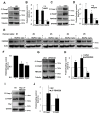
Figure 1
Figure 1
FBXO28 is reduced under diabetic conditions and its knockdown promotes β-cell apoptosis. INS-1E cells or isolated human islets were treated with (A,B) 22.2 mM glucose (HG), (E,F) the mixture of 22.2 mM glucose and 0.5 mM palmitate (HG/Pal), or (CF) pro-inflammatory cytokines (2 ng/mL recombinant human IL-1β, and 1000 U/mL IFN-γ; cyto) or transfected with either control scrambled siRNA (siScr) or siRNA specific to FBXO28 (siFBXO28, G,H) or with either control empty vector (EV)- or Myc-conjugated ∆F-FBXO28-overexpressing plasmids (I,J) for 2 (INS-1E) or 3 (human islets) days. Representative Western blots of cleaved caspase-3 (Cl Casp3), cleaved PARP (Cl PARP) and FBXO28 protein levels (A,C,E,G,I) and pooled densitometric analyses from at least three independent experiments (INS-1E; B,D,H,J) or six human islet preparations (F) are shown. GAPDH or Tubulin or Actin was analyzed to ensure equal protein loading. Data show means ± SEM. * p < 0.05 compared to control conditions.
Figure 2
Figure 2
FBXO28 overexpression protects from β-cell apoptosis under diabetic conditions. INS-1E cells were transfected with either control GFP- or FBXO28-overexpressing plasmids and treated with (A) 22.2 mM glucose (HG) or (C) pro-inflammatory cytokines (2 ng/mL recombinant human IL-1β, and 1000 U/mL IFN-γ; cyto) for 2 days. Representative Western blots of cleaved caspase-3 (Cl Casp3), cleaved PARP (Cl PARP) and Myc protein levels (A,C) and pooled densitometric analyses from at least three independent experiments (B,D) are shown. Tubulin was analyzed to ensure equal protein loading. Data show means ± SEM. * p < 0.05 compared to GFP transfected control conditions, ** p < 0.05 compared to GFP transfected diabetic (HG/Cyto) conditions.
Figure 3
Figure 3
FBXO28 does not regulate β-cell function in human islets. Freshly isolated human islets of nondiabetic organ donors were infected with LacZ control or FBXO28 adenoviruses (AF) or with Ad-GFP-shScr control or Ad-GFP-shFBXO28 (GL) for 2 days. (A,G) Insulin content analyzed after GSIS and normalized to whole islet protein. (B,H) Insulin secretion during 1 h-incubation with 2.8 mM (basal) and 16.7 mM glucose (stimulated), normalized to insulin content. (C,I) The insulin stimulatory index denotes the ratio of secreted insulin during 1 h-incubation with 16.7 mM and 2.8 mM glucose. (D,J) RT-PCR for NEUROD1, MAFA, PDX1, INS, GCK, NKX6.1, NKX2.2 and SlC2A2 normalized to Cyclophilin. FBXO28 mRNA (E,K) and protein (F,L) expression in human islets confirm successful FBXO28 overexpression (E,F) and downregulation (K,L). Pooled data are from at least four independent experiments from at least four different human islet donors. Data show means ± SEM. * p < 0.05 compared to Ad-LacZ (D,E) or Ad-shScr (J,K).
Figure 4
Figure 4
FBXO28 does not improve β-cell function in T2D human islets and FBXO28 mRNA expression is unchanged in T2D islets. (AD) Similar to non-diabetic human islets in Figure 3, freshly isolated human islets from patients with type 2 diabetes (T2D) were infected with LacZ control or FBXO28 adenoviruses for 1 day. (A) Insulin content analyzed after GSIS and normalized to whole islet protein; (B) Insulin secretion during 1 h-incubation with 2.8 mM (basal) and 16.7 mM glucose (stimulated), normalized to insulin content; (C) The insulin stimulatory index denotes the ratio of secreted insulin during 1 h-incubation with 16.7 mM and 2.8 mM glucose; (D) FBXO28 mRNA expression in human T2D islets; (E) RT-PCR for FBXO28 mRNA expression in human islets isolated from nondiabetic (n = 24) or individuals with T2D (n = 7), normalized to Cyclophilin. (AD) Pooled data are from five independent experiments from five different human islet donors with confirmed T2D. Data show means ± SEM. * p < 0.05 compared to Ad-LacZ.

Similar articles

Cited by

References

    1. Kurrer M.O., Pakala S.V., Hanson H.L., Katz J.D. β cell apoptosis in T cell-mediated autoimmune diabetes. Proc. Natl. Acad. Sci. USA. 1997;94:213–218. doi: 10.1073/pnas.94.1.213. - DOI - PMC - PubMed
    1. Butler A.E., Janson J., Bonner-Weir S., Ritzel R., Rizza R.A., Butler P.C. β-cell deficit and increased β-cell apoptosis in humans with type 2 diabetes. Diabetes. 2003;52:102–110. doi: 10.2337/diabetes.52.1.102. - DOI - PubMed
    1. Ardestani A., Paroni F., Azizi Z., Kaur S., Khobragade V., Yuan T., Frogne T., Tao W., Oberholzer J., Pattou F., et al. MST1 is a key regulator of β cell apoptosis and dysfunction in diabetes. Nat. Med. 2014;20:385–397. doi: 10.1038/nm.3482. - DOI - PMC - PubMed
    1. Talchai C., Xuan S., Lin H.V., Sussel L., Accili D. Pancreatic β cell dedifferentiation as a mechanism of diabetic β cell failure. Cell. 2012;150:1223–1234. doi: 10.1016/j.cell.2012.07.029. - DOI - PMC - PubMed
    1. Tiwari S., Roel C., Tanwir M., Wills R., Perianayagam N., Wang P., Fiaschi-Taesch N.M. Definition of a Skp2-c-Myc Pathway to Expand Human B-cells. Sci. Rep. 2016;6:28461. doi: 10.1038/srep28461. - DOI - PMC - PubMed

MeSH terms

LinkOut - more resources