

Surface Modification of Ti-35Nb-10Ta-1.5Fe by the Double Acid-Etching Process
- PMID: 29587427
- PMCID: PMC5951340
- DOI: 10.3390/ma11040494
Surface Modification of Ti-35Nb-10Ta-1.5Fe by the Double Acid-Etching Process
Abstract
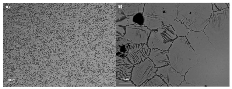
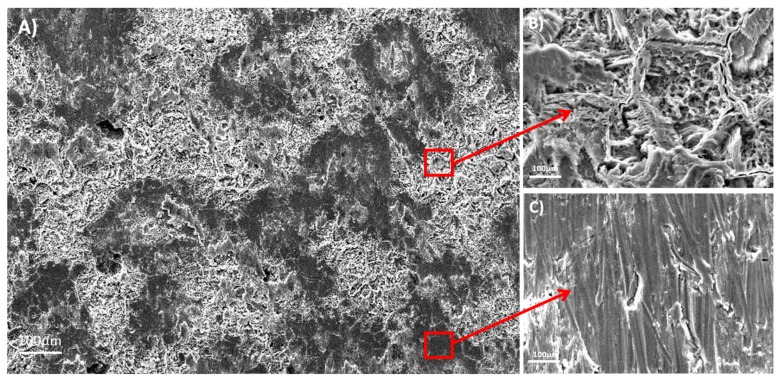
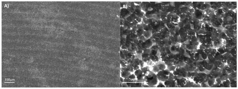
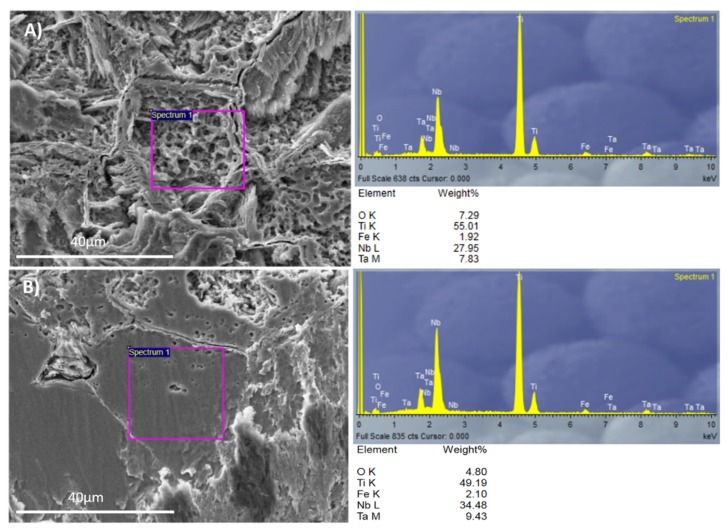
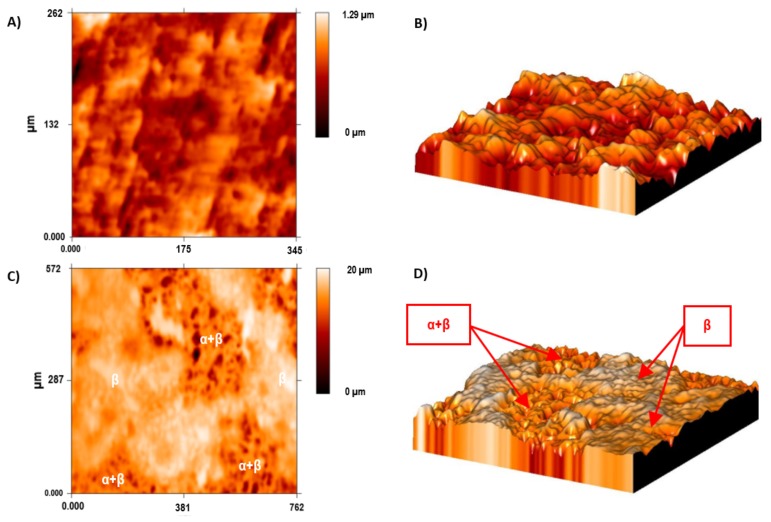
Surface topography and composition influence the osteoblastic proliferation and osseointegration rates, which favor the biomechanical stability of bone anchoring and implants. In recent years, beta titanium alloys have been developed, and are composed of biocompatible elements, have low elastic modulus, high corrosion resistance, and mechanical properties to improve the long performance behavior of biomaterials. In the present research, the influence of the acid-etching process was studied in Ti6Al4V ELI and Ti35Nb10Ta1.5Fe. Samples were etched in a two-step acid treatment. Surface roughness parameters were quantified under a confocal microscope, topography was studied by scanning electron microscopy, and surface composition was analyzed with energy dispersive X-ray spectroscopy. The results revealed that the two-step acid treatment changes the topography of the β alloy, increases the surface area, and changes the chemical composition of the surface. Two differentiated regions were identified in the Ti35Nb10Ta1.5Fe alloy after the acid-etching process: The α + β region with higher values of mean roughness due to the lower chemical resistance of this region; and the β region with lower values of roughness parameters.
Keywords: Ti-Nb-Ta-Fe; acid etching; beta alloy; surface roughness; titanium alloys; topography.
Conflict of interest statement
The authors declare no conflict of interest.
Figures
Similar articles
-
Investigations into Ti-(Nb,Ta)-Fe alloys for biomedical applications.Acta Biomater. 2016 Mar 1;32:336-347. doi: 10.1016/j.actbio.2015.12.010. Epub 2015 Dec 12. Acta Biomater. 2016. PMID: 26689463
-
Critical Role of Etching Parameters in the Evolution of Nano Micro SLA Surface on the Ti6Al4V Alloy Dental Implants.Materials (Basel). 2021 Oct 23;14(21):6344. doi: 10.3390/ma14216344. Materials (Basel). 2021. PMID: 34771869 Free PMC article.
-
Effect of surface treatment on bond strength of Ti-10Ta-10Nb to low-fusing porcelain.J Prosthet Dent. 2013 Feb;109(2):95-105. doi: 10.1016/S0022-3913(13)60023-2. J Prosthet Dent. 2013. PMID: 23395335
-
Commercially pure titanium (cp-Ti) versus titanium alloy (Ti6Al4V) materials as bone anchored implants - Is one truly better than the other?Mater Sci Eng C Mater Biol Appl. 2016 May;62:960-6. doi: 10.1016/j.msec.2016.01.032. Epub 2016 Jan 16. Mater Sci Eng C Mater Biol Appl. 2016. PMID: 26952502 Review.
-
Influence of surface electric charge of Ti implants on osteoblastic interaction: A systematic review.Saudi Dent J. 2022 Jul;34(5):335-345. doi: 10.1016/j.sdentj.2022.04.003. Epub 2022 Apr 21. Saudi Dent J. 2022. PMID: 35814840 Free PMC article. Review.
Cited by
-
Surface Modification Techniques to Produce Micro/Nano-scale Topographies on Ti-Based Implant Surfaces for Improved Osseointegration.Front Bioeng Biotechnol. 2022 Mar 25;10:835008. doi: 10.3389/fbioe.2022.835008. eCollection 2022. Front Bioeng Biotechnol. 2022. PMID: 35402405 Free PMC article. Review.
-
Laser-Based Ablation of Titanium-Graphite Composite for Dental Application.Materials (Basel). 2020 May 18;13(10):2312. doi: 10.3390/ma13102312. Materials (Basel). 2020. PMID: 32443423 Free PMC article.
-
Recent Advances and Prospects in β-type Titanium Alloys for Dental Implants Applications.ACS Biomater Sci Eng. 2024 Oct 14;10(10):6029-6060. doi: 10.1021/acsbiomaterials.4c00963. Epub 2024 Aug 30. ACS Biomater Sci Eng. 2024. PMID: 39215386 Free PMC article. Review.
References
-
- Lario-Femenía J., Amigó-Mata A., Vicente-Escuder A., Segovia-López F., Amigó-Borrás V. Desarrollo de las aleaciones de titanio y tratamientos superficiales para incrementar la vida útil de los implantes. Rev. Metal. 2016;52:e084. doi: 10.3989/revmetalm.084. - DOI
-
- Bjursten L.M., Rasmusson L., Oh S., Smith G.C., Brammer K.S., Jin S. Titanium dioxide nanotubes enhance bone bonding in vivo. J. Biomed. Mater. Res. Part A. 2010;92:1218–1224. - PubMed
-
- Davis J.R. Handbook of Materials for Medical Devices. ASM International; Geauga County, OH, USA: 2003.
-
- Kim E.S., Jeong Y.-H., Choe H.-C., Brantley W.A. Formation of titanium dioxide nanotubes on Ti-30Nb-xTa alloys by anodizing. Thin Solid Films. 2013;549:141–146. doi: 10.1016/j.tsf.2013.08.058. - DOI
LinkOut - more resources
Full Text Sources
Other Literature Sources
