Review
. 2018 Mar 26;6(2):38.
doi: 10.3390/biomedicines6020038.

NF-κB Activation in Lymphoid Malignancies: Genetics, Signaling, and Targeted Therapy

Affiliations
Review

NF-κB Activation in Lymphoid Malignancies: Genetics, Signaling, and Targeted Therapy

Paula Grondona et al. Biomedicines. .

Abstract

The NF-κB transcription factor family plays a crucial role in lymphocyte proliferation and survival. Consequently, aberrant NF-κB activation has been described in a variety of lymphoid malignancies, including diffuse large B-cell lymphoma, Hodgkin lymphoma, and adult T-cell leukemia. Several factors, such as persistent infections (e.g., with Helicobacter pylori), the pro-inflammatory microenvironment of the cancer, self-reactive immune receptors as well as genetic lesions altering the function of key signaling effectors, contribute to constitutive NF-κB activity in these malignancies. In this review, we will discuss the molecular consequences of recurrent genetic lesions affecting key regulators of NF-κB signaling. We will particularly focus on the oncogenic mechanisms by which these alterations drive deregulated NF-κB activity and thus promote the growth and survival of the malignant cells. As the concept of a targeted therapy based on the mutational status of the malignancy has been supported by several recent preclinical and clinical studies, further insight in the function of NF-κB modulators and in the molecular mechanisms governing aberrant NF-κB activation observed in lymphoid malignancies might lead to the development of additional treatment strategies and thus improve lymphoma therapy.

Keywords: CARD11; CARMA1; CD79; MyD88; NF-κB; leukemia; lymphoma.


Conflict of interest statement

The authors declare no conflict of interest.

Figures

Figure 1
Figure 1
Oncogenic MyD88 mutations activate the canonical NF-κB pathway. Activation of canonical NF-κB signaling can be induced by the triggering of members of the IL-1 receptor or the Toll-like receptor family, which localize to the cell surface or, in the case of TLR7 and TLR9, to the endosomal compartment. Ligand binding induces the recruitment of MyD88 to the activated receptor via its TIR domain and triggers subsequent downstream signaling. MyD88 oligomers nucleate the formation of the Myddosome which ultimately results in the activation of IRAKs and the recruitment of the E3 ubiquitin ligase TRAF6. TRAF6 in turn recruits and activates TAK1 which mediates activation of the IKK complex resulting in canonical NF-κB activity. The oncogenic variant MyD88L265P promotes spontaneous oligomerization and activation of downstream signaling independently of receptor stimulation. Mutant MyD88 is denoted with a red asterisk. DD, death domain; IL-1R, interleukin-1 receptor; IKK, IκB kinase; IRAK, IL-1R-associated kinase; MyD88, myeloid differentiation primary response protein 88; TAK1, TGFβ-activated kinase 1; TAB1/2, TAK1-binding protein 1/2; TIR; Toll/interleukin-1 receptor domain; TLR, Toll-like receptor; TRAF6, TNF receptor-associated factor 6.
Figure 2
Figure 2
Mechanisms of chronic active BCR signaling in lymphoid malignancies. (a) In the absence of its cognate antigen, proximal BCR signaling remains inactive, and thus the protein tyrosine kinase SYK is not recruited to the co-receptors CD79A and CD79B. The Src kinase LYN prevents hyperactivation of BCR signaling by initiation of a negative feedback loop involving CD22 phosphorylation and subsequent activation of the phosphatase SHP-1. Tonic BCR signaling promotes B-cell survival via the PI3K/AKT pathway in an antigen-independent manner; (b) Engagement of the BCR, for instance by self-antigens or pathogen-derived antigens, results in receptor clustering and the induction of an intracellular signaling cascade. The subsequent phosphorylation of tyrosine residues in the ITAM regions of CD79A/B by Src kinases (e.g., LYN) allows the recruitment of SYK which in turn phosphorylates the adapter protein BLNK and thus promotes the formation of a proximal signaling complex involving BTK and PLCγ2. While activation of the AKT signaling axis is achieved by the SYK-mediated phosphorylation of CD19 and subsequent recruitment of PI3K, PLCγ2 activity generates the second messengers DAG and IP3 the latter triggering the influx of Ca2+ into the cell. DAG and elevated Ca2+ levels activate PKCβ which induces activation of canonical NF-κB through the CBM signalosome. Whereas overexpression of SYK augments NF-κB activation in some lymphomas, mutations in the co-receptors CD79A/B prevent the internalization of activated BCRs and thus promote chronic BCR signaling. Proteins that are affected by recurrent genetic lesions in lymphoid malignancies are denoted with a red asterisk. BCR, B-cell receptor; BLNK, B-cell linker protein; BTK, Bruton’s tyrosine kinase; CBM, CARMA1/BCL10/MALT1; DAG, diacylglycerol; IP3, inositol 1,4,5-trisphosphate; ITAM, immunoreceptor tyrosine-based activation motif; PI3K, phosphoinositide 3-kinase; PKCβ, protein kinase Cβ; PLCγ2, phospholipase Cγ2; SHP-1, Src homology 2 domain phosphatase 1; SYK, spleen tyrosine kinase.
Figure 3
Figure 3
Increased activity of the CARMA1/BCL10/MALT1 signalosome drives constitutive NF-κB activation. Antigen-dependent triggering of the BCR results in the activation of PKCβ, which phosphorylates the scaffold protein CARMA1 at several residues within its inhibitory linker region. Activated CARMA1 in turn nucleates the formation of a fibrillary high-molecular weight signaling complex (CBM complex) comprising long BCL10 filaments and MALT1. Subsequently, the E3 ubiquitin ligase TRAF6 is recruited to the CBM complex via an interaction with MALT1 and catalyzes the K63-linked polyubiquitination of MALT1, BCL10, and itself. These K63-linked polyubiquitin chains as well as the linear polyubiquitin chains generated by the action of the LUBAC allow the recruitment and activation of the IKK complex. TAK1 which is recruited to the signaling complex via the ubiquitin binding domains of its accessory proteins TAB2 and TAB3 promotes IKK activation by phosphorylating IKKβ. IKK activity in turn results in the proteasomal degradation of IκBα and the subsequent nuclear translocation of transcriptionally active NF-κB heterodimers. In lymphoid malignancies, chronically active BCR signaling as well as gain-of-function mutations of the scaffold protein CARMA1 can lead to hyperactivation of the CBM signalosome and thus induce constitutive NF-κB activity. Genetic inactivation of A20, which negatively regulates IKK activity by deubiquitination of activated signaling effectors, augments NF-κB activation in several lymphoid cancers. Additionally, A20 can be proteolytically inactivated by the MALT1 protease, which is activated in the framework of the CBM complex. Expression of the oncogenic cIAP2-MALT1 fusion protein induces activation of canonical NF-κB in a TRAF6-dependent manner. The constitutive protease activity of cIAP2-MALT1 further promotes NF-κB activation by proteolytic inactivation of A20. Additionally, NIK cleavage by cIAP2-MALT1 also promotes non-canonical NF-κB activity. Proteins that are affected by recurrent genetic lesions in lymphoid malignancies are denoted with a red asterisk. BCL10, B-cell lymphoma 10; BCR, B-cell receptor; CARMA1, caspase recruitment domain membrane-associated guanylate kinase protein 1; cIAP2, cellular inhibitor of apoptosis protein 2; IκB, inhibitor of κB; IKK, IκB kinase; LUBAC, linear ubiquitin chain assembly complex; MALT1, mucosa-associated tissue lymphoma translocation protein 1; NIK, NF-κB inducing kinase; PKCβ, protein kinase Cβ; TAK1, TGFβ-activated kinase 1; TAB2/3, TAK1-binding protein 2/3; TRAF6, TNF receptor-associated factor 6.
Figure 4
Figure 4
Genetic lesions driving non-canonical NF-κB activation in lymphoid malignancies. (a) In unstimulated cells, TRAF3 recruits NIK to an E3 ubiquitin ligase complex comprising TRAF2 and cIAP1/2 which constantly marks NIK for proteasomal degradation by K48-linked polyubiquitination. The heterodimer of RelB and the p52 precursor protein p100 is sequestered in the cytoplasm; (b) Activated or mutated members of the TNF receptor family, such as CD40, BAFF-R, LTβR, and RANK or the EBV-encoded CD40 mimic LMP1, recruit the complex consisting of TRAF3, TRAF2, and cIAP1/2 to the cell membrane and induce the proteasomal degradation of TRAF3. In the absence of TRAF3, NIK is released from the inhibitory E3 ubiquitin ligase complex and accumulates in the cytoplasm where it subsequently phosphorylates and activates IKKα. In turn, IKKα phosphorylates the precursor protein p100, thus targeting it for processing by the proteasome. Proteasomal processing of p100 generates p52 and results in the nuclear translocation of transcriptionally active p52/RelB heterodimers. Enhanced expression of NIK due to copy number gains and loss-of-function mutations of TRAF2, TRAF3, or cIAP2 result in increased NIK protein levels driving non-canonical activation of NF-κB in lymphoid malignancies. Similarly, proteolytic cleavage by the constitutively active cIAP2-MALT1 fusion protein stabilizes NIK by removal of the TRAF3-binding site. Additionally, oncogenic MyD88 mutations can promote the activation of IKKα in a TAK1-dependent manner. In certain lymphomas aberrant activation of non-canonical NF-κB is governed by the expression of a truncated, constitutively active version of the precursor p100 lacking the inhibitory ankyrin repeats. Proteins that are affected by recurrent genetic lesions in lymphoid malignancies are denoted with a red asterisk. BAFF-R, B cell-activating factor receptor; cIAP1/2, cellular inhibitor of apoptosis protein 1/2; EBV, Epstein-Barr virus; IKKα, IκB kinase α; LMP1, latent membrane protein 1; LTβR, lymphotoxin β receptor; MALT1, mucosa-associated tissue lymphoma translocation protein 1; MyD88, myeloid differentiation primary response protein 88; NIK, NF-κB inducing kinase; RANK, receptor activator of NF-κB; TAK1, TGFβ-activated kinase 1; TRAF2/3, TNF receptor-associated factor 2/3.
Figure 5
Figure 5
Inactivating mutations in classical IκB proteins promote canonical NF-κB activation. (a) The canonical NF-κB pathway is induced by the ligand-dependent activation of a variety of receptors, such as antigen receptors, the IL-1 receptor, Toll-like receptors and members of the TNF receptor family. Stimulus-dependent activation of the IKK complex results in the phosphorylation and subsequent proteasomal degradation of IκBα and IκBε. This allows the nuclear translocation of transcriptionally active RelA/p50 or c-Rel/p50 heterodimers; (b) Genetic alterations resulting in the expression of non-functional truncated versions of IκBα and IκBε, which lack part of the ankyrin repeat domain and are thus unable to bind to the NF-κB transcription factors, promote constitutive NF-κB activation in a subset of lymphoid malignancies. Proteins that are affected by recurrent genetic lesions in lymphoid malignancies are denoted with a red asterisk. IκB, inhibitor of κB; IKK, IκB kinase.
Figure 6
Figure 6
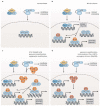
Abnormal expression of the atypical IκB proteins BCL3 and IκBζ promotes NF-κB activation in lymphoid malignancies. (a) In resting cells, DNA-bound homodimers of the NF-κB subunit p50 can be found in the nucleus due to constitutive processing of the p50 precursor p105; (b) Upon B-cell stimulation, the activated IKK complex induces both the generation of p50 by proteasomal processing of its precursor p105 and the proteasomal degradation of the inhibitor IκBα. This leads to the nuclear translocation of RelA/p50 heterodimers which drive the transcription of primary response genes, such as BCL3 and NFKBIZ. In turn, BCL3 and IκBζ modulate the secondary NF-κB response by binding to DNA-bound p50 homodimers in the nucleus; (c) The chromosomal translocation t(14;18)(q32;q13) juxtaposing BCL3 with the IGH locus, genomic amplifications, or epigenetic modifications result in enhanced expression of BCL3, which in turn increases the nuclear translocation of p50 and stabilizes DNA-bound p50 homodimers, thus augmenting the secondary NF-κB response; (d) Genomic amplification of the NFKBIZ locus as well as the expression of oncogenic MyD88L265P or the viral proteins Tax and LMP1 leads to increased expression of IκBζ in a subset of lymphoid tumors. IκBζ promotes the secondary NF-κB response through binding to p50 homodimers in the nucleus. BCL3, B-cell lymphoma 3; IGH, immunoglobulin heavy chain (locus); IκB, inhibitor of κB; IKK, IκB kinase; LMP1, latent membrane protein 1; MyD88, myeloid differentiation primary response protein 88.

References

    1. Liu F., Xia Y., Parker A.S., Verma I.M. IKK biology. Immunol. Rev. 2012;246:239–253. doi: 10.1111/j.1600-065X.2012.01107.x. - DOI - PMC - PubMed
    1. Ghosh S., Hayden M.S. New regulators of NF-κB in inflammation. Nat. Rev. Immunol. 2008;8:837–848. doi: 10.1038/nri2423. - DOI - PubMed
    1. Oeckinghaus A., Ghosh S. The NF-κB family of transcription factors and its regulation. Cold Spring Harb. Perspect. Biol. 2009;1:a000034. doi: 10.1101/cshperspect.a000034. - DOI - PMC - PubMed
    1. Sun S.C. The noncanonical NF-κB pathway. Immunol. Rev. 2012;246:125–140. doi: 10.1111/j.1600-065X.2011.01088.x. - DOI - PMC - PubMed
    1. Schuster M., Annemann M., Plaza-Sirvent C., Schmitz I. Atypical IκB proteins–nuclear modulators of NF-κB signaling. Cell Commun. Signal. 2013;11:23. doi: 10.1186/1478-811X-11-23. - DOI - PMC - PubMed

LinkOut - more resources