

Activated innate lymphoid cell populations accumulate in human tumour tissues
- PMID: 29587679
- PMCID: PMC5870240
- DOI: 10.1186/s12885-018-4262-4
Activated innate lymphoid cell populations accumulate in human tumour tissues
Abstract
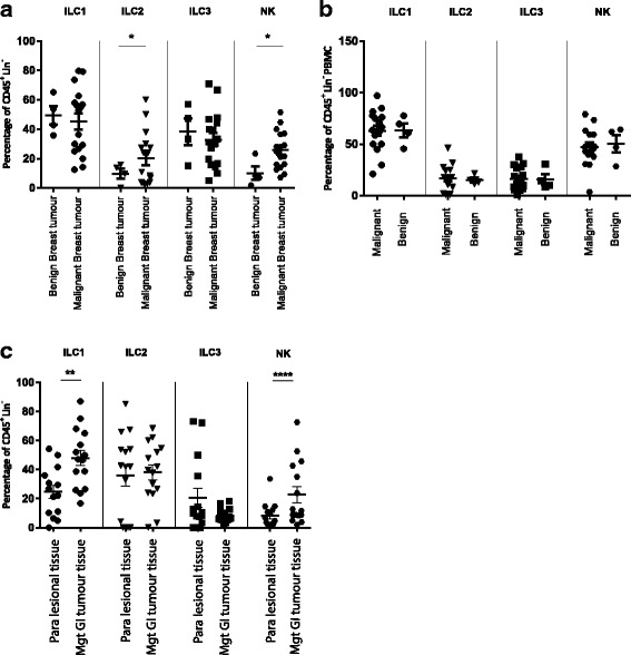
Background: Innate lymphoid cells (ILC) are part of a heterogeneous family of haematopoietic effector cells which lack re-arranged antigen-specific receptors. They promote host defense and contribute to tissue and metabolic homeostasis, wound healing and immune surveillance. Their role in human cancer immunity is less defined, and therefore we aimed to identify the frequency and phenotype of distinct ILC groups in various types of cancer.
Methods: Tissue samples and peripheral blood were collected from patients undergoing surgical resection of gastrointestinal and breast tumours. Single cell suspension of tumour tissue was immediately obtained following surgery using tumour dissociation.
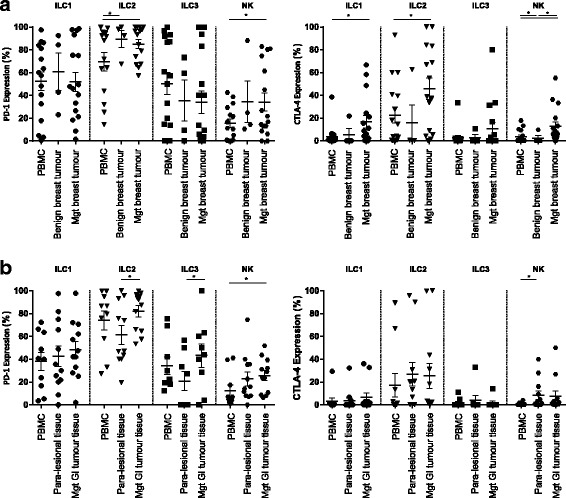
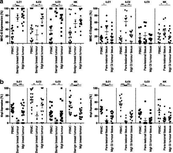
Results: We observed significantly higher frequencies of ILC2 (p value: 0.04) in malignant breast cancer tissue and significantly higher frequencies of group 1 ILC (p value: 0.001) in malignant gastrointestinal tumours. Tumour infiltrating ILC were found to show an activated phenotype with higher expression of MHC-II, KLRG1, early activation marker CD69 and CD44.
Conclusions: Activated innate lymphoid cells infiltrate tumours dependent on tumour type and location.
Keywords: Breast cancer; Gastrointestinal cancer; Immune checkpoint; Innate lymphoid cells.
Conflict of interest statement
Ethics approval and consent to participate
Written ethical approval was obtained from Xinjiang Tumour Hospital ethical committee and Oxford Tropical Research Ethics Committee (587–16) and London - City & East Research Ethics Committee (16/LO/1607). All participants gave informed written consent to participate.
Consent for publication
Not applicable.
Competing interests
The authors declare that they have no competing interests.
Publisher’s Note
Springer Nature remains neutral with regard to jurisdictional claims in published maps and institutional affiliations.
Figures

References
-
- Chow MT, Moller A, Smyth MJ. Inflammation and immune surveillance in cancer. Semin Cancer Biol. 2012;22(1):23–32. - PubMed
-
- Yang Q, et al. Antitumor activity of NK cells. Immunol Res. 2006;36(1–3):13–25. - PubMed
-
- Pages F, et al. Effector memory T cells, early metastasis, and survival in colorectal cancer. N Engl J Med. 2005;353(25):2654–2666. - PubMed
-
- Coulie PG, et al. Tumour antigens recognized by T lymphocytes: at the core of cancer immunotherapy. Nat Rev Cancer. 2014;14(2):135–146. - PubMed
Publication types
MeSH terms
Substances
Grants and funding
LinkOut - more resources
Full Text Sources
Other Literature Sources
Research Materials
Miscellaneous
