. 2018 Mar 27;49(1):31.
doi: 10.1186/s13567-018-0526-x.

Atrophy of primary lymphoid organs induced by Marek's disease virus during early infection is associated with increased apoptosis, inhibition of cell proliferation and a severe B-lymphopenia

Affiliations

Atrophy of primary lymphoid organs induced by Marek's disease virus during early infection is associated with increased apoptosis, inhibition of cell proliferation and a severe B-lymphopenia

Camille Berthault et al. Vet Res. .

Abstract

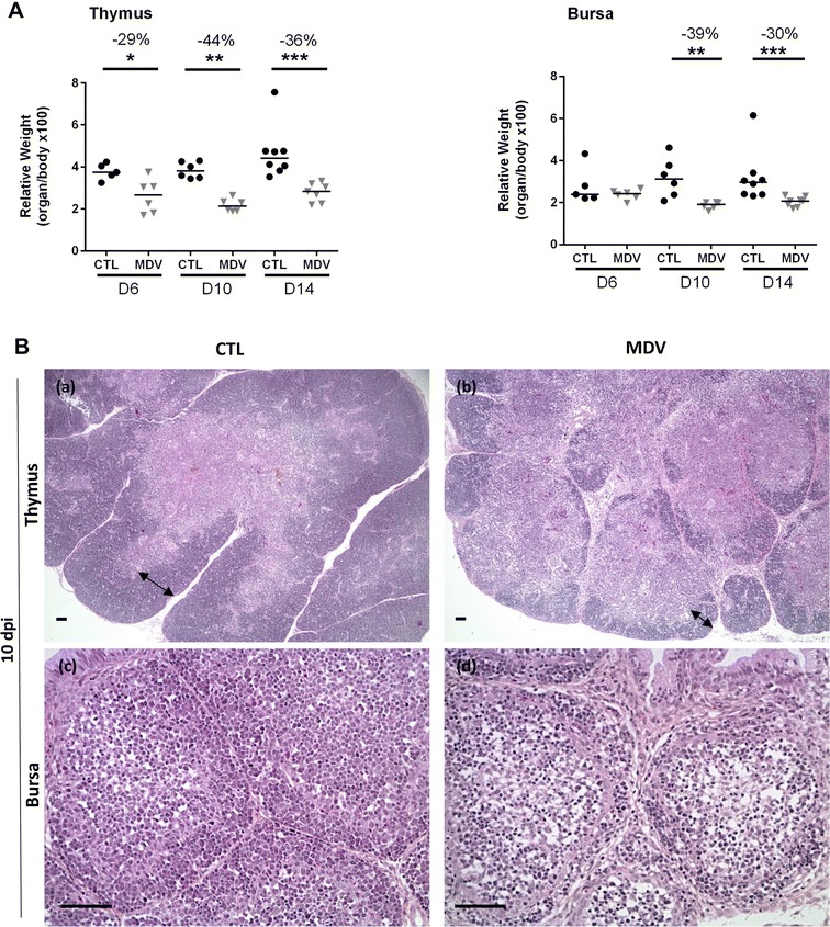
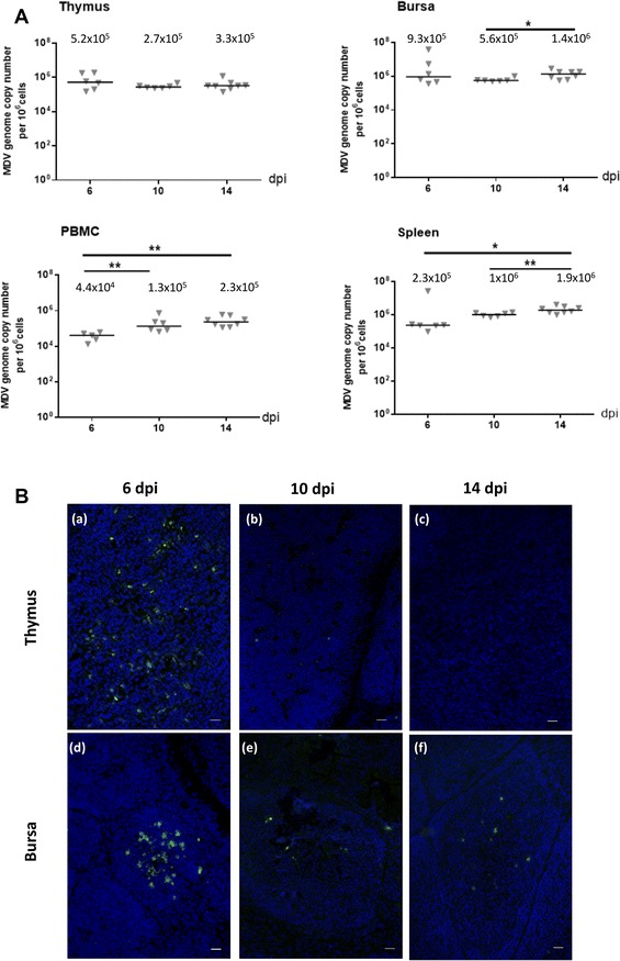
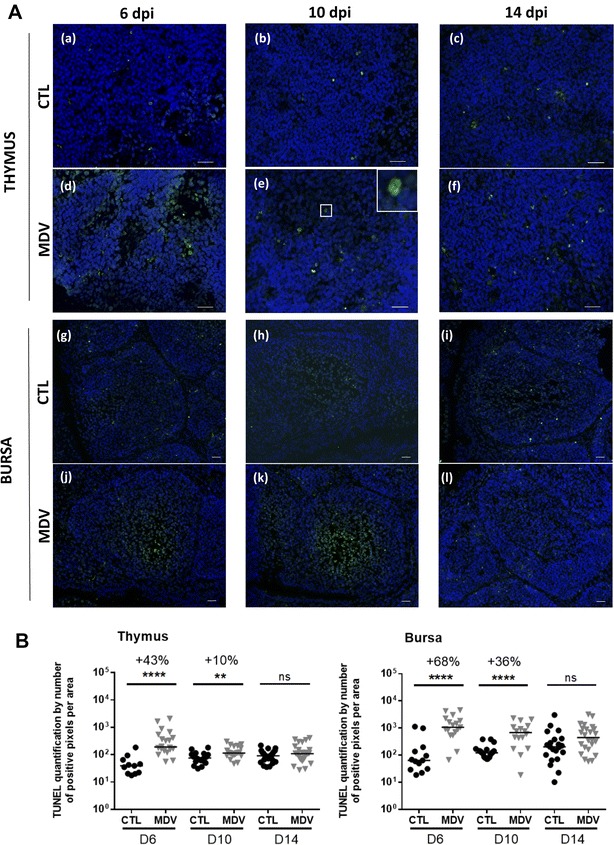
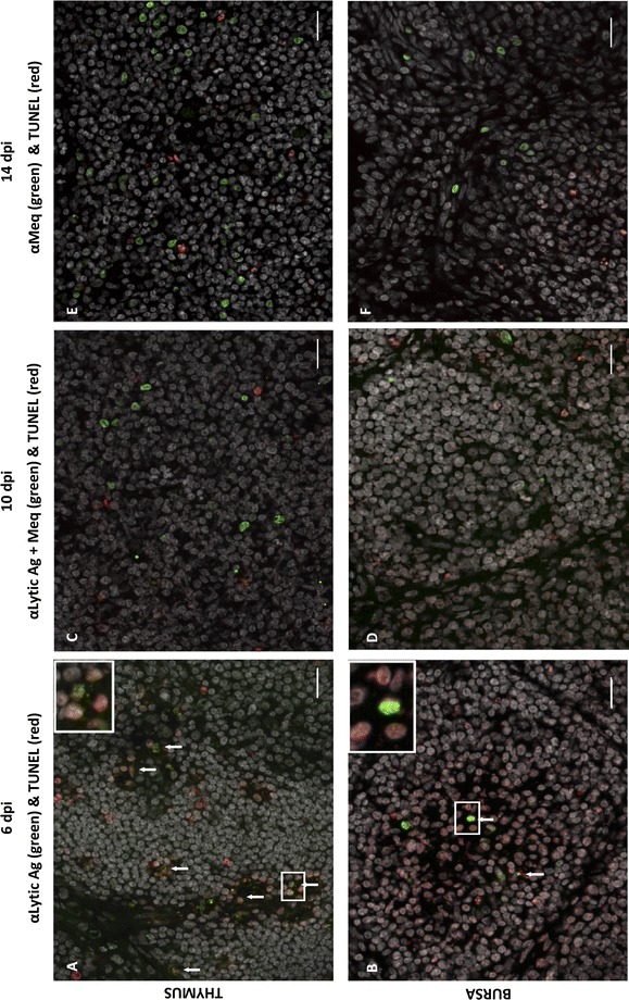
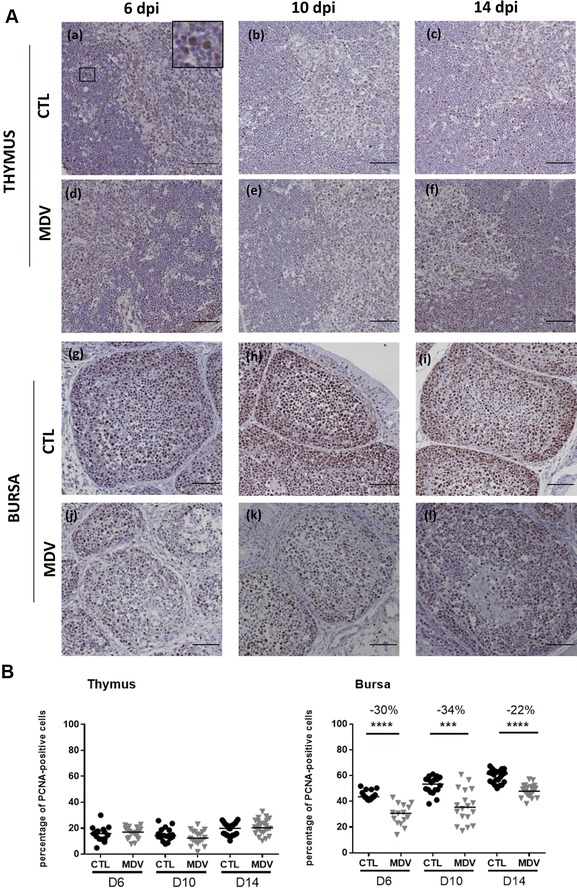
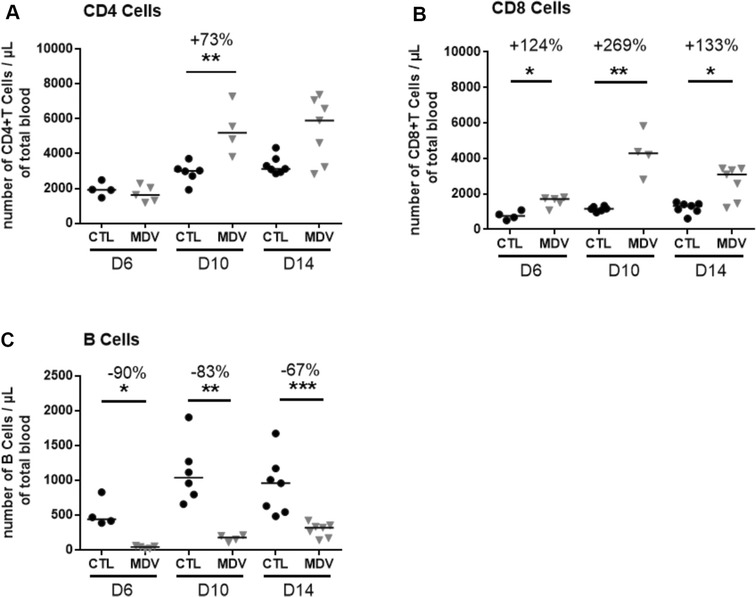
Marek's disease is a multi-faceted highly contagious disease affecting chickens caused by the Marek's disease alphaherpesvirus (MDV). MDV early infection induces a transient immunosuppression, which is associated with thymus and bursa of Fabricius atrophy. Little is known about the cellular processes involved in primary lymphoid organ atrophy. Here, by in situ TUNEL assay, we demonstrate that MDV infection results in a high level of apoptosis in the thymus and bursa of Fabricius, which is concomitant to the MDV lytic cycle. Interestingly, we observed that in the thymus most of the MDV infected cells at 6 days post-infection (dpi) were apoptotic, whereas in the bursa of Fabricius most of the apoptotic cells were uninfected suggesting that MDV triggers apoptosis by two different modes in these two primary lymphoid organs. In addition, a high decrease of cell proliferation was observed from 6 to 14 dpi in the bursa of Fabricius follicles, and not in the thymus. Finally, with an adapted absolute blood lymphocyte count, we demonstrate a major B-lymphopenia during the two 1st weeks of infection, and propose this method as a potent non-invasive tool to diagnose MDV bursa of Fabricius infection and atrophy. Our results demonstrate that the thymus and bursa of Fabricius atrophies are related to different cell mechanisms, with different temporalities, that affect infected and uninfected cells.


Figures

Figure 1
Figure 1
Thymus and bursa atrophy induced by the vvMDV RB-1B strain in White Leghorn B19/B19 chicks. A Organ weight/body weight ratios in CTL and MDV-infected chicks at 6, 10 and 14 dpi. The median is represented as a black line. Thymus and bursa relative weights were reduced significantly from 6 to 10 dpi respectively, in the MDV-infected group compared to the CTL group. B Histological structure of the thymus and the bursa at 10 dpi. Formalin-fixed tissues embedded in paraffin were sectioned and stained with HES. For the thymus, the cortex thickness (double-end arrow) was highly reduced in MDV-infected chicks (b) compared to controls (a). For the bursa, a massive cell depletion was observed in the medulla of the follicles in MDV-infected chicks (d) compared to the controls (c). Bar, 50 µm
Figure 2
Figure 2
MDV replication in lymphoid organs of infected-chicks at early time points. A MDV DNA loads in lymphoid organs of MDV-infected chicks. The MDV genome copy number per million cells was quantified at 6, 10 and 14 dpi using a Taqman real-time qPCR in thymus, bursa, PBMC and spleen. The viral loads were already high since 6 dpi in thymus, bursa, PBMC and spleen of all infected chicks and no significant or little further increase were detected at later time points. The median is represented as a black line. B Expression of lytic viral antigens in the thymus and the bursa. Cryosections of both thymus (a–c) and bursa (d–f) at 6, 10 and 14 dpi were stained with a cocktail of three mAb directed against MDV lytic antigens (VP22, ICP4, gB) (Green). Nuclei were stained with Hoescht 33342 dye (blue). MDV lytic antigens were detected in all birds in both primary lymphoid organs at 6 dpi; in 2/6 chicks for the thymus and 3/6 chicks for the bursa at 10 dpi and in 2/8 chicks for the bursa at 14 dpi (no detection in thymus at 14 dpi). In the bursa, MDV-infected cells in lytic cycle were predominantly located in the medulla at 6 dpi. Bar, 20 µm
Figure 3
Figure 3
MDV increases apoptosis in the thymus and the bursa at 6 and 10 dpi. A Cryosections of thymus (a–f) and bursa (g–l) harvested on CTL and MDV-infected birds at 6, 10 and 14 dpi were stained by TUNEL assay (green) and the nuclei counterstained with Hoescht 33342 dye (blue). TUNEL positive cells presented green nuclei, as shown enlarged (× 3) in the corner panel (e). Apoptosis was increased in the thymus of MDV-infected chicks compared to controls at 6 dpi, especially in the medulla. For the bursa, apoptosis was increased at 6 and 10 dpi, especially in the medulla. B Quantification of the green fluorescent signal on cryosections stained by TUNEL assay. This measurement was performed by area determination with FIJI software on three pictures per organ/chick/time. The median is represented with a black line. Apoptosis was highly enhanced at 6 dpi in the thymus and the bursa and to a lesser extend at 10 dpi compared to CTL. No differences in apoptosis were observed between infected- and CTL chicks at 14 dpi
Figure 4
Figure 4
Cells expressing MDV antigens are apoptotic in the thymus and rarely in the bursa. Cryosections of thymus (A, C, E) and bursa (B, D, F) were stained for MDV antigens (lytic: VP22, ICP4, gB and/or latent: Meq) (green) and apoptosis was detected by TUNEL assay (red). DNA content of the cells were stained with Hoescht 33342 dye (white). MDV antigens positive cells and apoptotic cells were detected in both organs at 6 dpi (A, B), 10 dpi (C, D) and 14 dpi (E, F). At 6 dpi, in the thymus, the majority of the cells expressing the lytic MDV-antigens were apoptotic, in contrast to the bursa. At 6 dpi, in the bursa, only rare double positive cells were detected (as shown in panel B, in the enlarged frame). In both organs, at 10 and 14 dpi, no co-localization between MDV antigens (including Meq) and TUNEL signal was observed. Bar, 20 µm
Figure 5
Figure 5
MDV inhibits cell proliferation in the bursa from 6 to 14 dpi but not in the thymus. A Proliferating cells were detected on both thymus (a–f) and bursa (g–l) of CTL and MDV-infected chicks at 6, 10 and 14 dpi by immunochemistry. For that, paraffin embedded sections were stained with a rabbit anti-cell nuclear antigen antibody (PCNA) revealed with EnVision anti-rabbit HRP kit after 3′diaminobenzidine (DAB) treatment (brown nuclei, as shown enlarged (×4) in the corner panel (a). The tissues were counterstained with Gill’s hematoxylin and the PCNA-negative nuclei appeared in blue. In the thymus, the number of PCNA-positive cells appeared to be equivalent in both groups of chicks. In the bursa, the total number of PCNA-positive cells was highly reduced in the infected group compared to the control group. PCNA-positive cells were mostly present in the bursal cortex of the control group. Bar, 50 µm. B Percentage of PCNA-positive cells in the bursa and the thymus at 6, 10 and 14 dpi. The median is represented with a black line. A significant reduction in the number of PCNA-positive cells was observed at each time point in the bursa, and not in the thymus
Figure 6
Figure 6
MDV infection induces a significant decrease in B-lymphocytes blood count at early time points. EDTA-blood of each chick was stained with anti-CD4-RPE, anti-CD8-FITC, anti-Bu1-PerCP-Cy5.5, anti-K1-RPE, and anti-CD45-APC in a no-lyse no-wash single-step procedure and analyzed by flow cytometry. Absolute number of T-CD4+ (A), T-CD8+ (B) and B lymphocytes (C) per µL of blood is given for most birds at each time point (6, 10 and 14 dpi). A few birds are missing because of partial blood clotting detected by cytometry. The median is represented with a black line. A significant increase was observed for T CD4+ cells at 10 dpi and for T CD8+ cells at all time points with a peak at day 10. Inversely, a significant decrease between 90 and 67% was observed for B-cells at 6, 10 and 14 dpi

References

    1. Morrow C, Fehler F. Marek’s disease: a worldwide problem. In: Davison F, Nair V, editors. Marek’s disease: an evolving problem. London: Elsevier Academic Press; 2004. pp. 49–61.
    1. Calnek BW. Lymphomagenesis in Marek’s disease. Avian Pathol. 1998;27:S54–S64. doi: 10.1080/03079459808419293. - DOI
    1. Schat KA. Marek’s disease immunosuppression. In: Davison F, Nair V, editors. Marek’s disease: an evolving problem. London: Elsevier Academic Press; 2004. pp. 17–31.
    1. Schat KA, Nair V. Marek’s disease. In: Saif YM, editor. Disease of poultry. Ames: Blackwell Publishing Ltd; 2008. pp. 452–514.
    1. Barrow AD, Burgess SC, Baigent SJ, Howes K, Nair VK. Infection of macrophages by a lymphotropic herpesvirus: a new tropism for Marek’s disease virus. J Gen Virol. 2003;84:2635–2645. doi: 10.1099/vir.0.19206-0. - DOI - PubMed

Publication types

LinkOut - more resources