Review
. 2018 Jul;28(7):523-540.
doi: 10.1016/j.tcb.2018.02.009. Epub 2018 Mar 24.

Endoplasmic Reticulum-Mitochondrial Contactology: Structure and Signaling Functions

Affiliations
Review

Endoplasmic Reticulum-Mitochondrial Contactology: Structure and Signaling Functions

György Csordás et al. Trends Cell Biol. 2018 Jul.

Abstract

Interorganellar contacts are increasingly recognized as central to the control of cellular behavior. These contacts, which typically involve a small fraction of the endomembrane surface, are local communication hubs that resemble synapses. We propose the term contactology to denote the analysis of interorganellar contacts. Endoplasmic reticulum (ER) contacts with mitochondria were recognized several decades ago; major roles in ion and lipid transfer, signaling, and membrane dynamics have been established, while others continue to emerge. The functional diversity of ER-mitochondrial (ER-mito) contacts is mirrored in their structural heterogeneity, with subspecialization likely supported by multiple, different linker-forming protein structures. The nanoscale size of the contacts has made studying their structure, function, and dynamics difficult. This review focuses on the structure of the ER-mito contacts, methods for studying them, and the roles of contacts in Ca2+ and reactive oxygen species (ROS) signaling.

Keywords: IP3 receptor; calcium ion; linkers; mitochondrion-associated membrane; ryanodine receptor; sarcoplasmic reticulum.


Figures

Figure 1
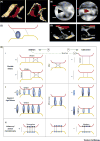
Figure 1. Shaping of the ER-mitochondrial contacts by linkers
A. Pseudo-colored electron-tomographic reconstructions of a 180 nm thick slice of a mitochondrion’s outer membrane (OMM, red) that forms contact with two ER sheets (yellow), from two different view angles. B. Zoomed-in image segments from the tomography slices used for A, showing electron-dense tethering structures (arrow heads) between the OMM and ER membranes (respectively indicated with the red and yellow lines). C. Tomographic reconstruction of the areas in B showing isodensity surfaces corresponding to ‘tethers’ (gray), OMM (red), ER (yellow) and ribosomes (blue ellipsoids). A–C are excerpts (from [9]) using specimen from DT40 chicken B cells. D. Simplified diagram for the ER-mito contact segments shown in C, with respective numbering. E. Diagrams illustrating how ER-mito contact dynamics is regulated by the membrane connecting elements. See text for details.
Figure 2
Figure 2. Diversity of distribution and interactions of ER-mito contact linker proteins
Venn diagram showing the subcellular distributions of a variety of ER-mito contact linker protein complexes between mitochondria, ER and cytosol; lines indicate protein-protein interactions. (The multimeric EMC is shown as a single entity for simplicity.)
Figure 3
Figure 3. Functions of the ER-mito contacts: membrane dynamics, signaling, lipid transfer
Top, schematized mitochondrion with a fission groove engaged to various contacts with sER tubules and a rER sheet. Below, on a schematized mitochondrial and interfacing ER segment, the molecular contributors of the main known functional interactions at ER-mito contacts are summarized. For bi/multipartite linkers the coupling domains are shown as blue clasps. Local Ca2+ fluxes are indicated with transparent red arrows while main mitochondria-derived ROS (·O2, H2O2) fluxes with transparent purple arrows. Abbreviations not yet introduced: PTP, permeability transition pore; Σ1, sigma-1 receptor; Roman I-V, respective respiratory chain complexes; TIMc/TOMc, protein translocase complexes of the IMM/OMM mediating mitochondrial protein import.

References

    1. Kerkhofs M, et al. Alterations in Ca2+ Signalling via ER-Mitochondria Contact Site Remodelling in Cancer. Advances in experimental medicine and biology. 2017;997:225–254. - PubMed
    1. Marchi S, et al. Endoplasmic Reticulum-Mitochondria Communication Through Ca2+ Signaling: The Importance of Mitochondria-Associated Membranes (MAMs) Advances in experimental medicine and biology. 2017;997:49–67. - PubMed
    1. Phillips MJ, Voeltz GK. Structure and function of ER membrane contact sites with other organelles. Nature reviews. Molecular cell biology. 2016;17:69–82. - PMC - PubMed
    1. Fernandez-Murray JP, McMaster CR. Lipid synthesis and membrane contact sites: a crossroads for cellular physiology. Journal of lipid research. 2016;57:1789–1805. - PMC - PubMed
    1. Paillusson S, et al. There's Something Wrong with my MAM; the ER-Mitochondria Axis and Neurodegenerative Diseases. Trends in neurosciences. 2016;39:146–157. - PMC - PubMed