Review
. 2019 Feb:86:102-111.
doi: 10.1016/j.semcdb.2018.03.017. Epub 2018 Mar 30.

Dynamics of E. coli single stranded DNA binding (SSB) protein-DNA complexes

Affiliations
Review

Dynamics of E. coli single stranded DNA binding (SSB) protein-DNA complexes

Edwin Antony et al. Semin Cell Dev Biol. 2019 Feb.

Abstract

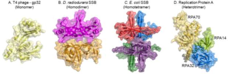
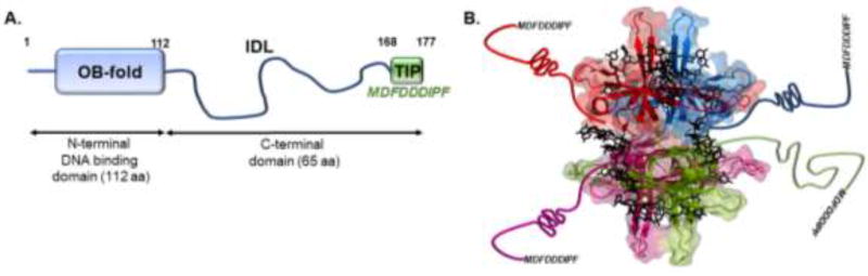
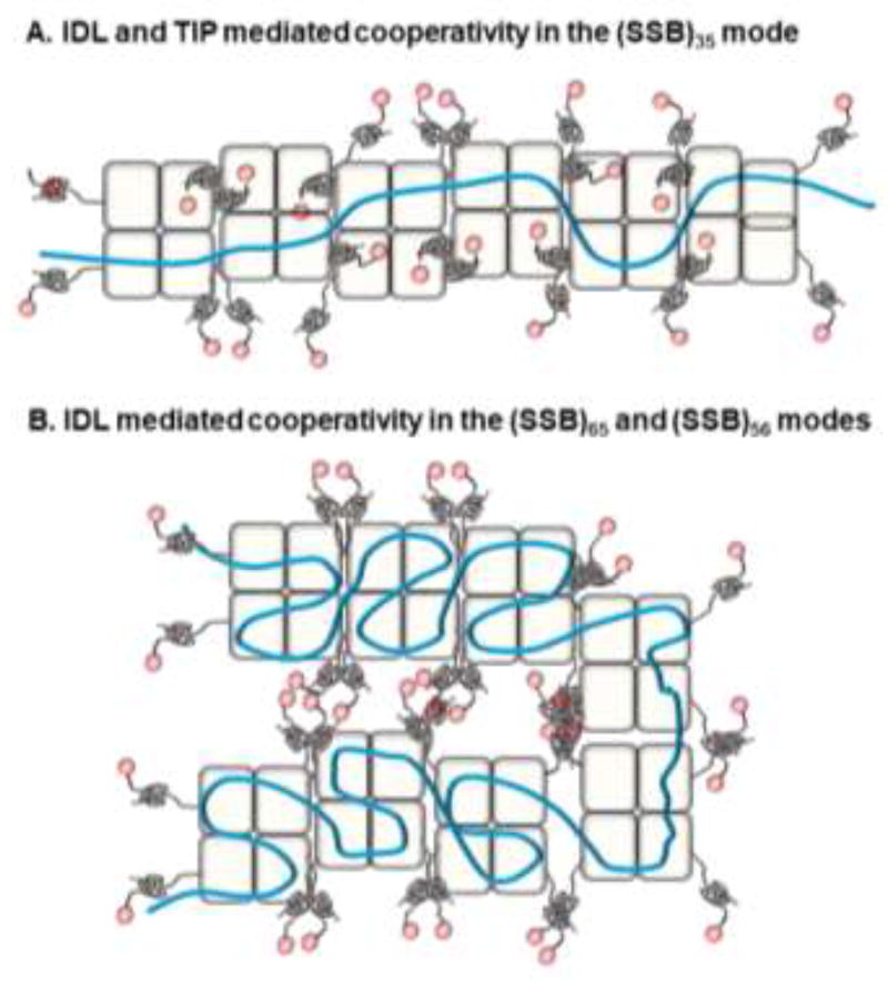
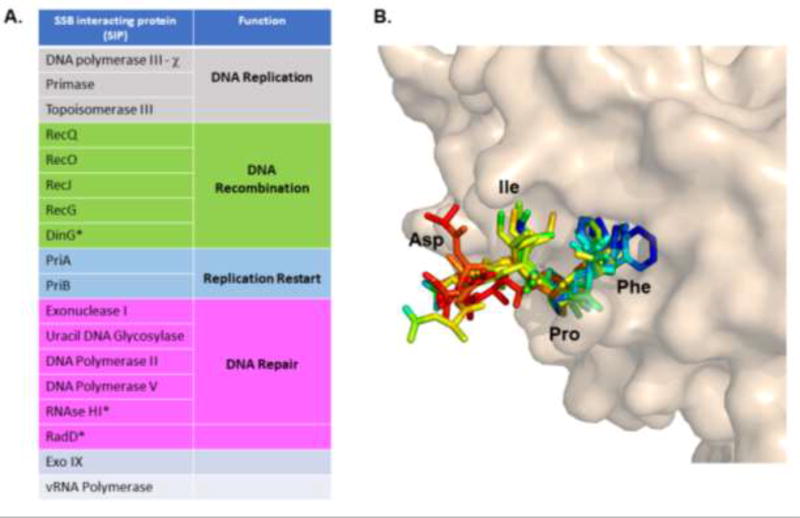
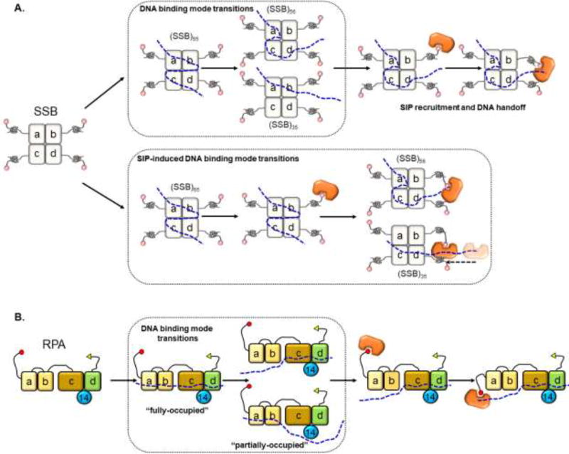
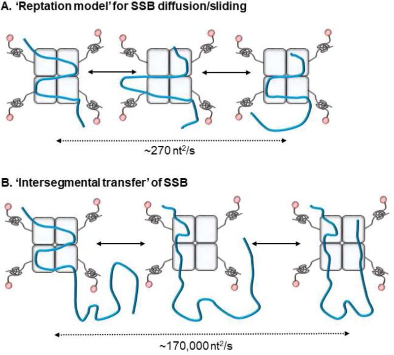
Single stranded DNA binding proteins (SSB) are essential to the cell as they stabilize transiently open single stranded DNA (ssDNA) intermediates, recruit appropriate DNA metabolism proteins, and coordinate fundamental processes such as replication, repair and recombination. Escherichia coli single stranded DNA binding protein (EcSSB) has long served as the prototype for the study of SSB function. The structure, functions, and DNA binding properties of EcSSB are well established: The protein is a stable homotetramer with each subunit possessing an N-terminal DNA binding core, a C-terminal protein-protein interaction tail, and an intervening intrinsically disordered linker (IDL). EcSSB wraps ssDNA in multiple DNA binding modes and can diffuse along DNA to remove secondary structures and remodel other protein-DNA complexes. This review provides an update on these features based on recent findings, with special emphasis on the functional and mechanistic relevance of the IDL and DNA binding modes.

Keywords: Diffusion; Intersegmental transfer; RPA; SSB; ssDNA.


Figures

Figure 1
Figure 1. Subunit composition of single strand DNA binding proteins
Crystal structures of SSB proteins from various organisms and their respective oligomeric states are depicted. Structures were generated from the following PDB IDs: 1GPC, 3UDG, 1EYG and 4GNX.
Figure 2
Figure 2. Architecture of EcSSB
A) Schematic of the DNA binding oligonucleotide/oligosaccharide-binding (OB) domain, the C-terminal TIP and the intervening intrinsically disordered loop (IDL) of EcSSB. B) Crystal structure of EcSSB (cartoon) bound to ssDNA (sticks; 1EYG) is shown with each subunit colored. The IDLs are shown extending away from the DNA binding core and the sequence of the TIP are denoted.
Figure 3
Figure 3. Models of IDL and TIP mediated cooperativity in EcSSB
A) Cooperative binding of SSB tetramers in the (SSB)35 mode is shown. Proposed interactions between the IDLs of neighboring tetramers along with TIP interactions with free ssDNA binding regions in the OB-domains are denoted. B) Similar cooperative binding to ssDNA in the (SSB)65 and (SSB)56 modes are proposed to be facilitated through interactions between IDLs of multiple tetramers.
Figure 4
Figure 4. SSB interactions with SSB interacting proteins (SIPs)
A) An updated list of the SIPs identified to date are categorized according to their cellular function. The asterisks denote newly identified SIPs. B) The crystal structure of exonuclease I in complex with the terminal four residues in the EcSSB TIP (PDB ID: 3C94) is shown. Similar TIP regions from multiple SIP-TIP peptide structures were aligned and shown here colored according to conformational flexibility (B-factors); red and blue denote extremes of high and low flexibility, respectively. The Ile-Pro-Phe residues adopt similar conformations in all these structures, whereas the Asp residue situated away from the active site can adopt multiple conformations.
Figure 5
Figure 5. DNA binding mode transitions in SSB
A) SSB can spontaneously transition between the (SSB)35, (SSB)56, and (SSB)65 binding modes, and the transiently open ssDNA allow binding of SIPs. A model where SIPs facilitate transitions between binding modes is also depicted. B) The four-OB domains in RPA that are primarily responsible for ssDNA binding are shown. DNA binding domains (DBD) a, b and c reside in RPA70 and are connected by flexible linkers. DBD c, d and the RPA14 subunit form the trimerization core. RPA is also proposed to transition between multiple binding modes enabling the binding of RPA interacting proteins (orange) to ssDNA vacated by one or more DBDs.
Figure 6
Figure 6. Diffusion of SSB
A) The reptation model for EcSSB diffusion/sliding is shown where segment(s) of ssDNA-OB domain interactions are perturbed allowing another OB-domain to bind to the free DNA. This enables the entire tetramer to shift or diffuse along the DNA lattice. B) On long ssDNA, SSB achieves rapid diffusion using principles of direct transfer where transiently dissociated ssDNA are replaced by DNA from a distant location. The respective rates for the two processes are denoted.

References

    1. Sigal N, Delius H, Kornberg T, Gefter ML, Alberts B. A DNA-unwinding protein isolated from Escherichia coli: its interaction with DNA and with DNA polymerases. Proceedings of the National Academy of Sciences of the United States of America. 1972;69:3537–41. - PMC - PubMed
    1. Molineux IJ, Gefter ML. Properties of the Escherichia coli DNA-binding (unwinding) protein interaction with nucleolytic enzymes and DNA. Journal of molecular biology. 1975;98:811–25. - PubMed
    1. Weiner JH, Bertsch LL, Kornberg A. The deoxyribonucleic acid unwinding protein of Escherichia coli. Properties and functions in replication. The Journal of biological chemistry. 1975;250:1972–80. - PubMed
    1. Chrysogelos S, Griffith J. Escherichia coli single-strand binding protein organizes single-stranded DNA in nucleosome-like units. Proceedings of the National Academy of Sciences of the United States of America. 1982;79:5803–7. - PMC - PubMed
    1. Schneider RJ, Wetmur JG. Kinetics of transfer of Escherichia coli single strand deoxyribonucleic acid binding protein between single-stranded deoxyribonucleic acid molecules. Biochemistry. 1982;21:608–15. - PubMed

Publication types

MeSH terms

LinkOut - more resources