. 2018 Aug 28;138(9):898-912.
doi: 10.1161/CIRCULATIONAHA.117.032636.

Cholesterol Efflux Pathways Suppress Inflammasome Activation, NETosis, and Atherogenesis

Affiliations

Cholesterol Efflux Pathways Suppress Inflammasome Activation, NETosis, and Atherogenesis

Marit Westerterp et al. Circulation. .

Abstract

Background: The CANTOS trial (Canakinumab Antiinflammatory Thrombosis Outcome Study) showed that antagonism of interleukin (IL)-1β reduces coronary heart disease in patients with a previous myocardial infarction and evidence of systemic inflammation, indicating that pathways required for IL-1β secretion increase cardiovascular risk. IL-1β and IL-18 are produced via the NLRP3 inflammasome in myeloid cells in response to cholesterol accumulation, but mechanisms linking NLRP3 inflammasome activation to atherogenesis are unclear. The cholesterol transporters ATP binding cassette A1 and G1 (ABCA1/G1) mediate cholesterol efflux to high-density lipoprotein, and Abca1/g1 deficiency in myeloid cells leads to cholesterol accumulation.

Methods: To interrogate mechanisms connecting inflammasome activation with atherogenesis, we used mice with myeloid Abca1/g1 deficiency and concomitant deficiency of the inflammasome components Nlrp3 or Caspase-1/11. Bone marrow from these mice was transplanted into Ldlr-/- recipients, which were fed a Western-type diet.

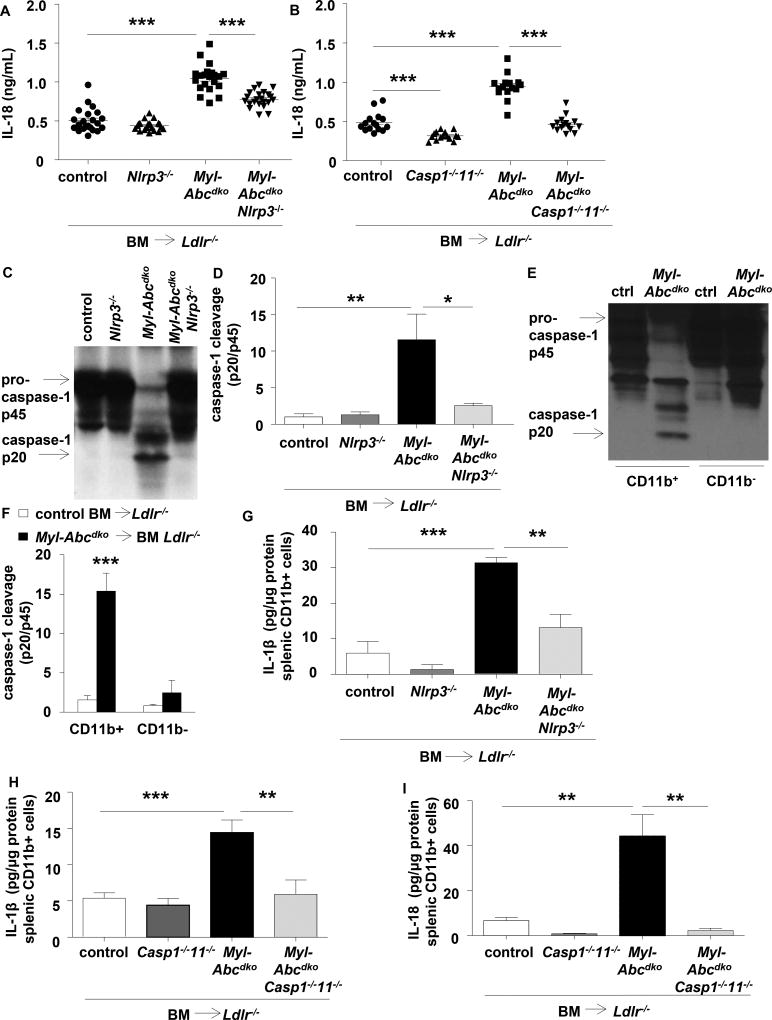
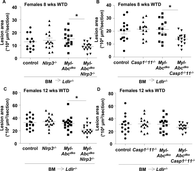
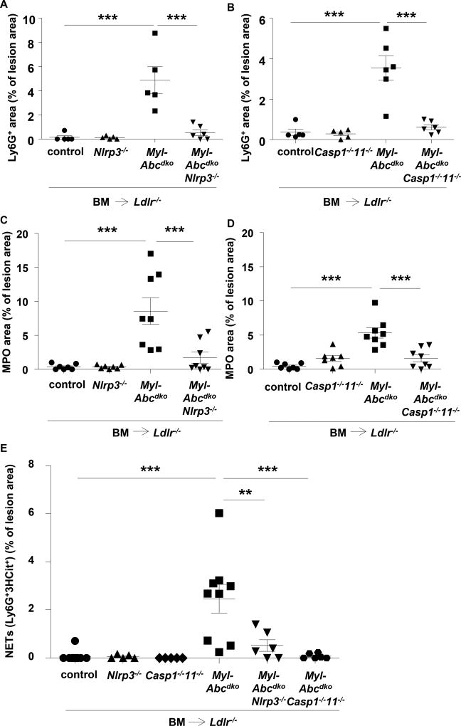
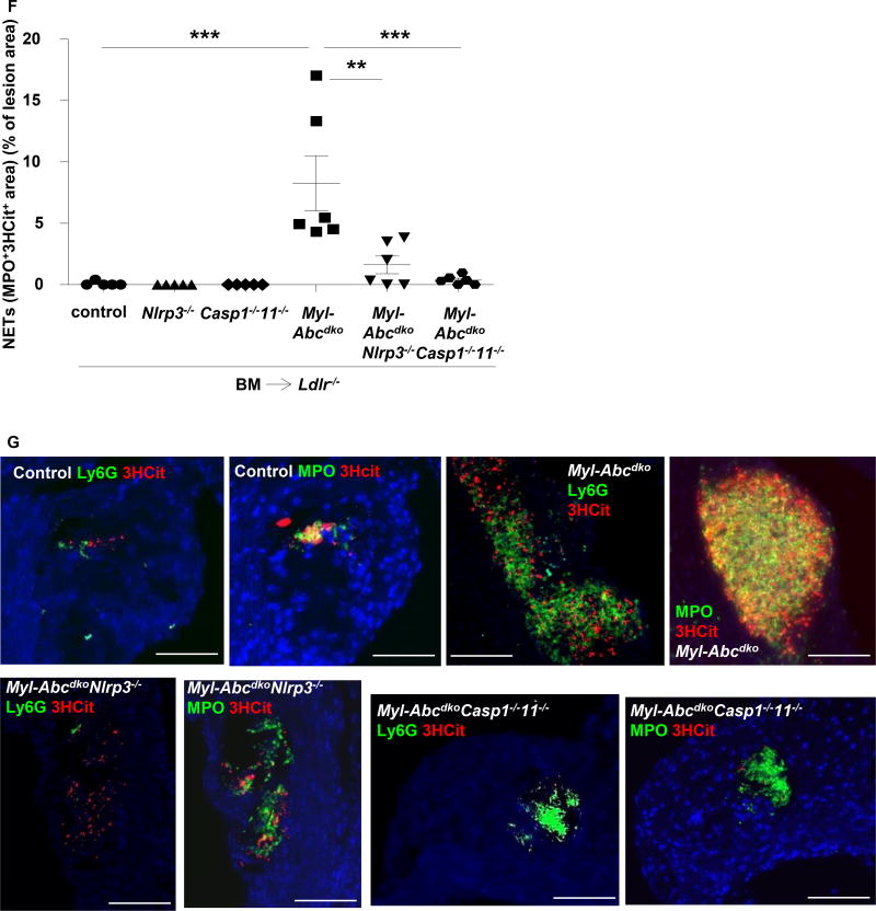
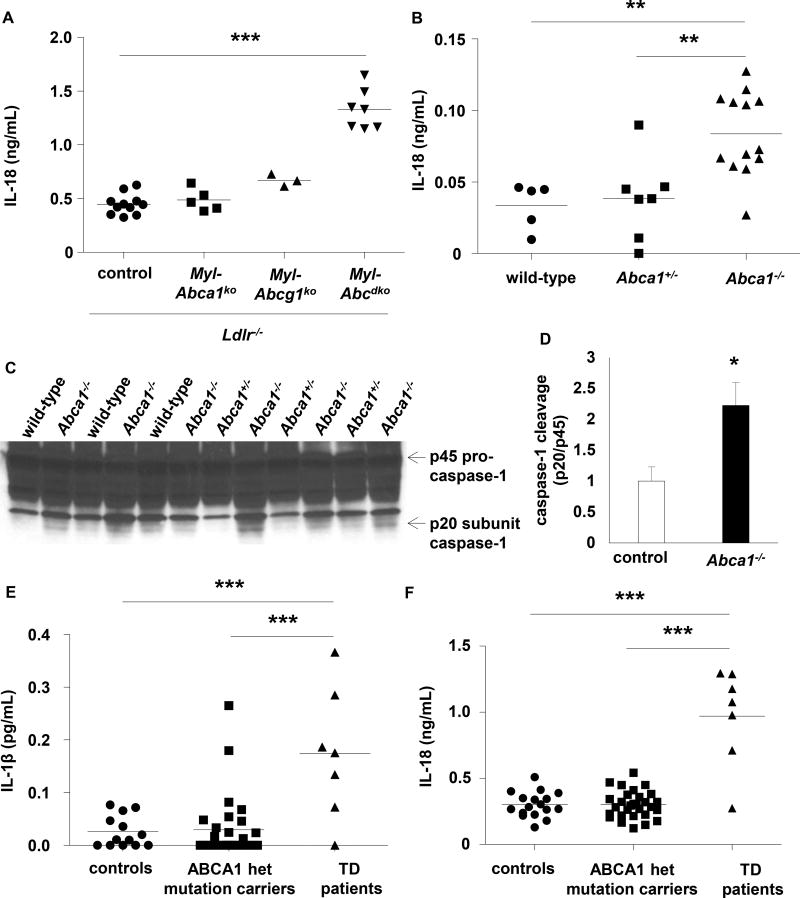
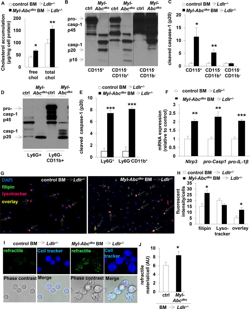
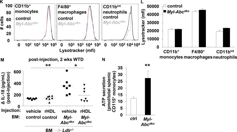
Results: Myeloid Abca1/g1 deficiency increased plasma IL-18 levels in Ldlr-/- mice and induced IL-1β and IL-18 secretion in splenocytes, which was reversed by Nlrp3 or Caspase-1/11 deficiency, indicating activation of the NLRP3 inflammasome. Nlrp3 or Caspase-1/11 deficiency decreased atherosclerotic lesion size in myeloid Abca1/g1-deficient Ldlr-/- mice. Myeloid Abca1/g1 deficiency enhanced caspase-1 cleavage not only in splenic monocytes and macrophages, but also in neutrophils, and dramatically enhanced neutrophil accumulation and neutrophil extracellular trap formation in atherosclerotic plaques, with reversal by Nlrp3 or Caspase-1/11 deficiency, suggesting that inflammasome activation promotes neutrophil recruitment and neutrophil extracellular trap formation in atherosclerotic plaques. These effects appeared to be indirectly mediated by systemic inflammation leading to activation and accumulation of neutrophils in plaques. Myeloid Abca1/g1 deficiency also activated the noncanonical inflammasome, causing increased susceptibility to lipopolysaccharide-induced mortality. Patients with Tangier disease, who carry loss-of-function mutations in ABCA1 and have increased myeloid cholesterol content, showed a marked increase in plasma IL-1β and IL-18 levels.

Conclusions: Cholesterol accumulation in myeloid cells activates the NLRP3 inflammasome, which enhances neutrophil accumulation and neutrophil extracellular trap formation in atherosclerotic plaques. Patients with Tangier disease, who have increased myeloid cholesterol content, showed markers of inflammasome activation, suggesting human relevance.

Keywords: ATP-binding cassette transporters; atherosclerosis; cholesterol, HDL; extracellular traps; inflammasomes.


Conflict of interest statement

Conflict of Interest Disclosures

A.R.T. reports being a consultant to Amgen, CSL, Staten Biotech, and Dalcor. The other authors report no conflicts of interest.

Figures

Figure 1
Figure 1. Myeloid Abca1/g1 deficiency enhances NLRP3 inflammasome activation in Ldlr−/− mice
Ldlr−/− mice were transplanted with bone marrow (BM) from control, Myl-Abcdko, Nlrp3−/−, Myl-AbcdkoNlrp3−/−, Caspase1−/−11−/−, or Myl-AbcdkoCaspase1−/−11−/− mice and fed Western type diet (WTD) for 4 weeks. Genotypes of BM donors are indicated on the graphs. (A–B) Plasma IL-18 levels (n=12–20; each datapoint represents one mouse). (C–D) Caspase-1 cleavage and quantification in total splenocytes, and (E–F) splenic CD11b+ and CD11b cells (n=6). (G–I) IL-1β and IL-18 secretion in splenic CD11b+ cells. (n=6). (F). *P<0.05, **P<0.01, ***P<0.001 by t-test, or (A–B, D, G–I) one-way ANOVA with Bonferroni post-test.
Figure 2
Figure 2. Inflammasome activation accelerates atherosclerosis in myeloid Abca1/g1 deficiency
Ldlr−/− mice were transplanted with BM from control, Myl-Abcdko, Nlrp3−/−, Myl-AbcdkoNlrp3−/−, Caspase1−/−11−/−, or Myl-AbcdkoCaspase1−/−11−/− mice, and fed WTD for 8 or 12 weeks, as indicated. (A–D) Sections were stained with haematoxylin-eosin (H&E) and atherosclerotic lesion area was measured at the level of the aortic root (n=13–17 mice per group). In (A–D) each datapoint represents a single mouse. (A–D) *P<0.05 by one-way ANOVA with Bonferroni post-test.
Figure 3
Figure 3. Inflammasome activation promotes neutrophil accumulation and neutrophil extracellular trap formation in atherosclerotic lesions of myeloid Abca1/g1 deficient mice
Ldlr−/− mice were transplanted with BM from control, Myl-Abcdko, Nlrp3−/−, Myl-AbcdkoNlrp3−/−, Caspase1−/−11−/−, or Myl-AbcdkoCaspase1−/−11−/− mice, and fed WTD for 8 weeks. (A–D) In atherosclerotic lesions of mice, neutrophils were stained using Ly6G (A–B) and activated neutrophils using myeloperoxidase (MPO) (C–D) Ly6G+ and MPO+ percentage of lesion size was quantified (n=5–8 mice per group). (E–F) Lesions were also stained for citrullinated histones 2, 8, and 17 (3HCit). To assess neutrophil extracellular traps (NETs), the overlap of Ly6G and 3HCit (E) or MPO and 3HCit (F) was quantified (n=5–7 mice per group). (G) Representative examples are shown. In (A–F) each datapoint represents one mouse. **P<0.01, ***P<0.001 by one-way ANOVA with Bonferroni post-test. Scale bars represent 100 µm.
Figure 4
Figure 4. Mice with whole body Abca1 deficiency and humans with homozygous loss-of-function mutations for ABCA1 show signs of inflammasome activation
(A) Ldlr−/− mice were transplanted with BM from control, Myl-Abca1ko, Myl-Abcg1ko, or Myl-Abcdko mice, and fed WTD for 4 weeks. Plasma IL-18 levels were assessed. (B–D) Wild-type, Abca1+/−, and Abca1−/− mice were fed WTD for 4 weeks and plasma IL-18 levels (B) and caspase-1 cleavage in total splenocytes (C–D) were assessed. In (A–B), each datapoint represents one mouse. Controls in (D) represent wild-type and Abca1+/− mice. (E–F) IL-1β (E) and IL-18 (F) levels were measured in plasma of Tangier Disease (TD) patients carrying a homozygous loss-of-function mutation for ABCA1, and gender and age matched heterozygous ABCA1 mutation carriers and controls. Each datapoint represents one patient or control. *P<0.05, **P<0.01, ***P<0.001 by t-test (D) or one-way ANOVA with Bonferroni post-test (A–B) or a mixed effects model with random intercepts taking into account data-clustering due to family members (E–F).
Figure 5
Figure 5. Inflammasome activation in myeloid Abca1/g1 deficiency is cholesterol-dependent
Ldlr−/− mice were transplanted with BM from control or Myl-Abcdko mice, as indicated, and fed WTD for 8 weeks unless indicated otherwise. Genotypes of BM donors are indicated on the graphs. (A) Splenic Ly6G+ cells were isolated, lipids extracted, and free cholesterol and total cholesterol levels were assessed. (B–E) Caspase-1 cleavage and quantification in (B–C) splenic CD115+, CD115CD11b+, and CD115CD11b cells, and (D–E) splenic Ly6G+, and Ly6GCD11b+ cells. (F) Inflammasome priming in splenic CD11b+ cells. (n=6). (G–H) Splenic CD115+CD11b+ cells were isolated and stained with lysotracker and filipin. Arrowheads indicate overlay between lysotracker and filipin (G). Representative examples (G) and quantification (H) are shown. (I–J) Refractile material was assessed in total splenocytes using confocal microscopy. Representative examples (I) and quantification (J) are shown. (K–L) Lysotracker staining was assessed using flow cytometry in CD11b+ monocytes, F4/80+ macrophages, and CD11bint neutrophils, gated as in Suppl Fig 8A. (M) Mice were fed WTD for 1 week, injected with reconstituted HDL (rHDL; CSL-111; 120 mg/kg) and the increase in plasma IL-18 levels was assessed at 1 week after injection (n=7 mice per group; each datapoint represents one mouse). (N) ATP secretion from total splenic CD115+ monocytes was assessed using luciferase assay. n=6 (A, C, E–F, H, J, L, N), or n=7 (M) mice per group. (A, C, E–F, H, J, L, N) *P<0.05, **P<0.01, ***P<0.001 by t-test, or (M) one-way ANOVA with Bonferroni post-test.
Figure 6
Figure 6. Myeloid Abca1/g1 deficiency activates the non-canonical inflammasome
(A–D) Ldlr−/− mice were transplanted with BM from control, Myl-Abcdko, Nlrp3−/−, or Myl-AbcdkoNlrp3−/− mice (n=6 per group), and fed WTD for 8 weeks. Genotypes of BM donors are indicated on the graph. Caspase-11 cleavage was assessed in splenic CD11b+ cells (A–B) or Ly6G+ and Ly6GCD11b+ cells (C–D) using Western blot, and quantified (B, D). (B) *P<0.05, **P<0.01, ***P<0.001 by one-way ANOVA with Bonferroni post-test or (D) t-test. (E) Mice of the indicated genotypes (n=8 per group) were fed a chow diet and injected with LPS at 8 weeks of age (20 mg/kg; i.p.). Mortality was assessed every 6 hours over a 48 h time period. aP<0.05 control compared to Caspase1−/−11−/−; bP<0.05 Nlrp3−/− compared to Caspase1−/−11−/−; cP<0.01 control compared to Myl-Abcdko; dP<0.01 Nlrp3−/− compared to Myl-AbcdkoNlrp3−/−; eP<0.001 Myl-Abcdko compared to Myl-AbcdkoCaspase1−/−11−/−; fP<0.001 Myl-AbcdkoNlrp3−/− compared to Myl-AbcdkoCaspase1−/−11−/−, Log-rank (Mantel-Cox) test. Reported significance values are nominal and uncorrected.

Comment in

References

    1. Ridker PM, Everett BM, Thuren T, MacFadyen JG, Chang WH, Ballantyne C, Fonseca F, Nicolau J, Koenig W, Anker SD, Kastelein JJP, Cornel JH, Pais P, Pella D, Genest J, Cifkova R, Lorenzatti A, Forster T, Kobalava Z, Vida-Simiti L, Flather M, Shimokawa H, Ogawa H, Dellborg M, Rossi PRF, Troquay RPT, Libby P, Glynn RJ, Group CT. Antiinflammatory therapy with canakinumab for atherosclerotic disease. N Engl J Med. 2017;377:1119–1131. - PubMed
    1. Jaiswal S, Fontanillas P, Flannick J, Manning A, Grauman PV, Mar BG, Lindsley RC, Mermel CH, Burtt N, Chavez A, Higgins JM, Moltchanov V, Kuo FC, Kluk MJ, Henderson B, Kinnunen L, Koistinen HA, Ladenvall C, Getz G, Correa A, Banahan BF, Gabriel S, Kathiresan S, Stringham HM, McCarthy MI, Boehnke M, Tuomilehto J, Haiman C, Groop L, Atzmon G, Wilson JG, Neuberg D, Altshuler D, Ebert BL. Age-related clonal hematopoiesis associated with adverse outcomes. N Engl J Med. 2014;371:2488–2498. - PMC - PubMed
    1. Fuster JJ, MacLauchlan S, Zuriaga MA, Polackal MN, Ostriker AC, Chakraborty R, Wu CL, Sano S, Muralidharan S, Rius C, Vuong J, Jacob S, Muralidhar V, Robertson AA, Cooper MA, Andres V, Hirschi KK, Martin KA, Walsh K. Clonal hematopoiesis associated with tet2 deficiency accelerates atherosclerosis development in mice. Science. 2017;355:842–847. - PMC - PubMed
    1. Duewell P, Kono H, Rayner KJ, Sirois CM, Vladimer G, Bauernfeind FG, Abela GS, Franchi L, Nunez G, Schnurr M, Espevik T, Lien E, Fitzgerald KA, Rock KL, Moore KJ, Wright SD, Hornung V, Latz E. Nlrp3 inflammasomes are required for atherogenesis and activated by cholesterol crystals. Nature. 2010;464:1357–1361. - PMC - PubMed
    1. Elhage R, Jawien J, Rudling M, Ljunggren HG, Takeda K, Akira S, Bayard F, Hansson GK. Reduced atherosclerosis in interleukin-18 deficient apolipoprotein e-knockout mice. Cardiovasc Res. 2003;59:234–240. - PubMed

Publication types

MeSH terms