. 2018 May 11;293(19):7376-7386.
doi: 10.1074/jbc.RA117.000358. Epub 2018 Mar 27.

A cassette of basic amino acids in histone H2B regulates nucleosome dynamics and access to DNA damage

Affiliations

A cassette of basic amino acids in histone H2B regulates nucleosome dynamics and access to DNA damage

Yesenia Rodriguez et al. J Biol Chem. .

Abstract

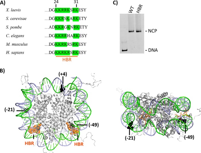
Nucleosome dynamics, such as spontaneous DNA unwrapping, are postulated to have a critical role in regulating the access of DNA repair machinery to DNA lesions within nucleosomes. However, the specific histone domains that regulate nucleosome dynamics and the impact of such changes in intrinsic nucleosome dynamics on DNA repair are not well understood. Previous studies identified a highly conserved region in the N-terminal tail of histone H2B known as the histone H2Brepression (or HBR) domain, which has a significant influence on gene expression, chromatin assembly, and DNA damage formation and repair. However, the molecular mechanism(s) that may account for these observations are limited. In this study, we characterized the stability and dynamics of ΔHBR mutant nucleosome core particles (NCPs) in vitro by restriction enzyme accessibility (REA), FRET, and temperature-induced sliding of histone octamers. Our results indicate that ΔHBR-NCPs are more dynamic, with a larger steady-state fraction of the NCP population occupying the unwrapped state than for WT-NCPs. Additionally, ΔHBR-histone octamers are more susceptible to temperature-induced sliding on DNA than WT histone octamers. Furthermore, we show that the activity of base excision repair enzymes at uracil lesions and single nucleotide gaps is enhanced in a site-specific manner in ΔHBR-NCPs. This enhanced activity correlates well with regions exhibiting increased DNA unwrapping. Finally, removal of the HBR domain is not sufficient to completely alleviate the structural constraints imposed by histone octamers on the activity of base excision repair enzymes.

Keywords: DNA repair; HBR domain; base excision repair (BER); chromatin; glycosylase; histone H2B repression; histone modification; nucleosome; polymerase beta.


Conflict of interest statement

The authors declare that they have no conflicts of interest with the contents of this article

Figures

Figure 1.
Figure 1.
The HBR domain in histone H2B. A, HBR sequence alignment from various species was adapted from Parra et al. (27). As can be seen, the HBR domain (e.g. residues 24–31 in X. laevis, 30–37 in S. cerevisiae, and 27–37 in Homo sapiens) is highly conserved among species and is predominantly comprised of positively charged amino acids (50, 51). B, in the left figure, PDB code 1KX5 was modified to highlight the structural location of the HBR domain (orange spheres) relative to BER lesions (black spheres). A different view with the HBR domain displayed as sticks is shown on the right. For the lesions (uracil or single-nucleotide gap), the number in parentheses indicates the number of nucleotides away from the pseudo 2-fold axis of symmetry called the dyad, toward the 5′ end (+) or (−) toward the 3′ end of the damaged strand. All lesions are inwardly oriented or occluded, with their phosphate backbone facing the histone octamer. Lesions at −49 and +4 located in the I strand (green) and −21 in the J strand (blue). C, representative 6% native gel illustrating reconstitution efficiency of DNA with recombinant X. laevis WT and HBR (Δ24–31) octamers.
Figure 2.
Figure 2.
Restriction enzyme accessibility assay on nucleosomal DNA containing WT (unmodified) or ΔHBR (H2BΔ24–31 amino acids) histone octamers. A, schematic of the nucleosome core particle showing the approximate locations of two restriction sites relative to the dyad, where the number in parentheses indicates the cleavage site (base number) on the I strand toward the 5′ end from the dyad. The samples were incubated with either HaeIII or MspI, where indicated (+) for 2 h at 37 °C. In B, the substrate contained a uracil at −21 in B and at +4 in C. Error bars represent standard deviation of the means for at least three independent experiments. Two-tailed, unpaired t tests were performed, and asterisks indicate the level of significance with p values of 0.0001 and 0.004 for MspI and HaeIII, respectively, in B and 0.002 in C.
Figure 3.
Figure 3.
Nucleosome dynamics of WT and ΔHBR NCPs as assessed by FRET. A, the crystal structure PDB code 1KX5 was modified to highlight the structural location of fluorophore FRET pairs, where red indicates the positioning of cy5 (acceptor) and green the positioning of cy3 (donor). For end-labeled NCPs, a Cy3-labeled base was inserted at position 6, and a Cy5-labeled base at position 81 in the 601 sequence, as described previously (37). For HBR-label NCPs, Cy3 and Cy5 fluorophores were similarly located at base position 46 from the 5′ end of the I chain and base position 25 from the 5′ end of the J chain, respectively. Internal-label fluorophores were located at position 33 from the 5′ end of the I chain (Cy3) and position 34 from the 5′ end of the J chain (Cy5). The panels on the left show emission spectra for WT and ΔHBR NCPs with FRET pairs located at the end-label (B), HBR-label (D), and internal-label (F) positions. The panels on the right show salt-induced nucleosome disassembly/unwrapping for WT and ΔHBR NCPs monitored by FRET using end-labeled (C), HBR-label (E), and internal-label (G) NCPs. Salt concentrations were adjusted to the appropriate final concentration with 5 m NaCl and allowed to equilibrate for 30 min at room temperature. Emission spectra at the salt concentrations shown were normalized to the Cy5 signal excited at 615 nm. The FRET efficiency at each salt concentration was normalized to the corresponding low salt efficiency to allow comparison of the salt-induced FRET decrease between WT and HBR NCPs.
Figure 4.
Figure 4.
Thermal repositioning of WT and ΔHBR NCPs. A, schematic diagram of NCPs reconstituted with the 256-bp 601 DNA positioning sequence. Strongest NCP positions are indicated by solid ellipses, and weaker positions are indicated by dashed lines. B, representative 5% nondenaturing polyacrylamide gels of WT and ΔHBR NCPs reconstituted with the 256-bp 601 DNA positioning sequence. C, quantification of the fraction of DNA in each NCP structure (center or side) or as naked DNA at different temperatures. Error bars represent ± S.D. of the mean of at least three independent experiments.
Figure 5.
Figure 5.
Effect of HBR deletion on uracil removal by UDG-APE1. 601 DNA, containing a single uracil at the specified locations, was 5′-end–labeled with either [γ-32P]ATP or Cy3 and annealed with the corresponding undamaged complementary strand. Substrates were reconstituted with WT or ΔHBR histone octamers, and DNA cleavage reactions were performed at 37 °C for the specified time with UDG (30 nm) and APE1 (10 nm). Cleavage activity was measured on denaturing gels (see insets), as described under “Experimental procedures.” The data points represent the means ± S.D. of at least three independent experiments.
Figure 6.
Figure 6.
Effect of HBR deletion on DNA synthesis by Pol β. WT and ΔHBR NCPs containing a single-nucleotide gap, located at positions denoted at the top of each panel, were incubated with 100 nm Pol β, a 5-fold molar excess relative to nucleosomal DNA. Extension products were separated on 8% polyacrylamide denaturing sequencing gels. Representative gels (imbedded within each chart) are shown with an arrow denoting the extension product band. Data points represent the means ± S.D. of at least three independent experiments.
Figure 7.
Figure 7.
Model describing how HBR domain regulates NCP dynamics and DNA repair. In WT NCPs (upper panel), the HBR domain (depicted in orange) represses spontaneous nucleosomal DNA unwrapping, which limits access of repair enzymes (green oval) to DNA lesions (cyan) in nucleosomes. In HBR deletion mutants (lower panel), spontaneous nucleosomal DNA unwrapping is increased, which facilitates repair of DNA lesions. We hypothesize that in vivo HBR PTMs may also facilitate nucleosomal DNA unwrapping.

References

    1. Luger K., Mäder A. W., Richmond R. K., Sargent D. F., and Richmond T. J. (1997) Crystal structure of the nucleosome core particle at 2.8 Å resolution. Nature 389, 251–260 10.1038/38444 - DOI - PubMed
    1. Guintini L., Charton R., Peyresaubes F., Thoma F., and Conconi A. (2015) Nucleosome positioning, nucleotide excision repair and photoreactivation in Saccharomyces cerevisiae. DNA Repair (Amst.) 36, 98–104 10.1016/j.dnarep.2015.09.012 - DOI - PubMed
    1. Rodriguez Y., Hinz J. M., and Smerdon M. J. (2015) Accessing DNA damage in chromatin: preparing the chromatin landscape for base excision repair. DNA Repair (Amst.) 32, 113–119 10.1016/j.dnarep.2015.04.021 - DOI - PMC - PubMed
    1. McGinty R. K., and Tan S. (2015) Nucleosome structure and function. Chem. Rev. 115, 2255–2273 10.1021/cr500373h - DOI - PMC - PubMed
    1. Hodges A. J., Gallegos I. J., Laughery M. F., Meas R., Tran L., and Wyrick J. J. (2015) Histone sprocket arginine residues are important for gene expression, DNA repair, and cell viability in Saccharomyces cerevisiae. Genetics 200, 795–806 10.1534/genetics.115.175885 - DOI - PMC - PubMed

Publication types

LinkOut - more resources