. 2018 Mar 23:11:79.
doi: 10.1186/s13068-018-1063-6. eCollection 2018.

A fast and sensitive activity assay for lytic polysaccharide monooxygenase

Affiliations

A fast and sensitive activity assay for lytic polysaccharide monooxygenase

Erik Breslmayr et al. Biotechnol Biofuels. .

Abstract

Background: Lytic polysaccharide monooxygenases (LPMO) release a spectrum of cleavage products from their polymeric substrates cellulose, hemicellulose, or chitin. The correct identification and quantitation of these released products is the basis of MS/HPLC-based detection methods for LPMO activity. The duration, effort, and intricate analysis allow only specialized laboratories to measure LPMO activity in day-to-day work. A spectrophotometric assay will simplify the screening for LPMO in culture supernatants, help monitor recombinant LPMO expression and purification, and support enzyme characterization.

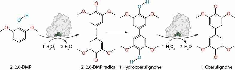
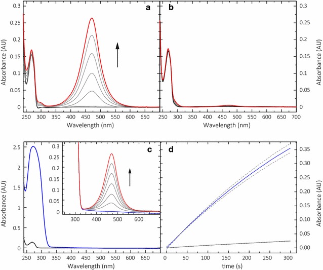
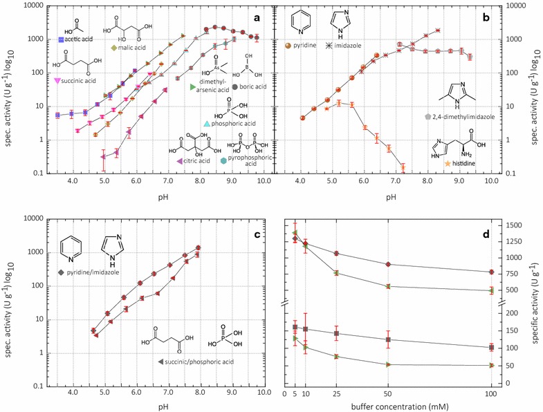
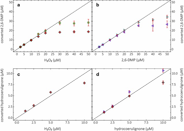
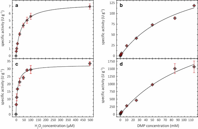
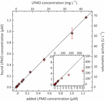
Results: Based on a newly discovered peroxidase activity of LPMO, we propose a fast, robust, and sensitive spectrophotometric activity assay using 2,6-dimethoxyphenol (2,6-DMP) and H2O2. The fast enzymatic assay (300 s) consists of 1 mM 2,6-DMP as chromogenic substrate, 100 µM H2O2 as cosubstrate, and an adequate activity of LPMO in a suitable buffer. The high molar absorption coefficient of the formed product coerulignone (ε469 = 53,200 M-1 cm-1) makes the assay sensitive and allows reliable activity measurements of LPMO in concentrations of approx. 0.5-50 mg L-1.

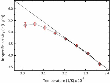
Conclusions: The activity assay based on 2,6-DMP detects a novel peroxidase activity of LPMO. This activity can be accurately measured and used for enzyme screening, production, and purification, and can also be applied to study binding constants or thermal stability. However, the assay has to be used with care in crude extracts, because other enzymes such as laccase or peroxidase will interfere with the assay. We also want to stress that the peroxidase activity is a homogeneous reaction with soluble substrates and should not be correlated to heterogeneous LPMO activity on polymeric substrates.

Keywords: 2,6-Dimethoxyphenol; Activity assay; Biomass degradation; Hydrogen peroxide; Lytic polysaccharide monooxygenase; Peroxidase activity.


Figures

Fig. 1
Fig. 1
2,6-DMP oxidation. LPMO catalyzes the oxidation of 2,6-DMP to the corresponding phenoxy radical at the expense of H2O2. The active-site Cu(II) is reduced by 2,6-DMP which generates the 2,6-DMP radical. Two formed 2,6-DMP radicals dimerize rapidly to hydrocoerulignone, which again is quickly converted to coerulignone by LPMO. The stoichiometry of the peroxidase reaction is 1:1
Fig. 2
Fig. 2
Conversion of 200 µM 2,6-DMP and hydrocoerulignone by 2 µM NcLPMO9C in 100 mM sodium succinate/phosphate buffer, pH 6.0. a Reaction of 2,6-DMP in the presence of 100 µM H2O2 and 250 µM dissolved O2, and b in the presence of 250 µM O2 only, followed for 5000 s. Time frequency between each trace is 1000 s. c Spectra of 2,6-DMP (black line) and hydrocoerulignone (blue line), 200 µM each. Inset demonstrates the conversion of hydrocoerulignone; note that the occurring peak is similar to a, followed for 300 s. Time frequency between each trace is 50 s. d Time course of the NcLPMO9C (0.3 µM)-catalyzed conversion of 1 mM 2,6-DMP (black line) and 1 mM hydrocoerulignone (blue line) measured at 469 nm in the presence of 100 µM H2O2 in 100 mM sodium succinate/phosphate buffer, pH 6.0. The data are expressed as mean values (± SD), from three independent repeats
Fig. 3
Fig. 3
pH-dependent activity of NcLPMO9C (0.10 – 3.75 µM; 30 µM for histidine chloride buffer) in various buffers with 25 mM 2,6-DMP and 100 µM H2O2. a 100 mM sodium carboxylate, arsenate, phosphate, and borate buffers. b 100 mM imidazole or pyridine chloride buffers. c Anionic and cationic mixed buffers spanning a broader pH range. d Sensitivity of the NcLPMO9C-catalyzed reaction to ionic strength in the cationic broad range buffer (squares) and the anionic broad range buffer (triangles) at pH 6.0 (lower traces) and 7.5 (higher traces). The data are expressed as mean values (± SD), from three independent repeats
Fig. 4
Fig. 4
Reaction stoichiometry. Calculated concentrations of converted 2,6-DMP (top graphs) or hydrocoerulignone (bottom graphs) after oxidation with NcLPMO9C, laccase, and horseradish peroxidase. Experiments with setting different H2O2 concentrations (left graphs) or setting different concentrations of 2,6-DMP or hydrocoerulignone (right graphs). a Linear increase of converted 2,6-DMP at pH 7.5 until 20 µM for NcLPMO9C (black diamonds) or 30 µM for horseradish peroxidase (green circle). b Linear increase of converted 2,6-DMP at pH 6.0 (blue triangle) and pH 7.5 (cyan triangles) until 30 µM for laccase without addition of H2O2. c Linear increase of converted hydrocoerulignone at pH 6.0 and d at pH 7.5 until 5 µM for NcLPMO9C (black diamonds; with addition of 100 µM H2O2) and pH 6.0 for laccase (blue triangle; without addition of H2O2). The data are expressed as mean values (± SD), from at least three independent repeats. Linear range was taken to calculate the molar absorption coefficient for coerulignone (ε469 = 53,200 M−1 cm−1). Dashed line represents the ideal 1:1 stoichiometry for the 2,6-DMP:H2O2 ratio based on the molar absorption coefficient
Fig. 5
Fig. 5
Michaelis–Menten kinetics at pH 6.0 (a, b) and pH 7.5 (c, d). For H2O2 kinetics: graphs with different H2O2 concentrations (a, c) and 1 mM 2,6-DMP. For 2,6-DMP kinetics: graphs with different 2,6-DMP concentrations (b, d) and 100 µM H2O2
Fig. 6
Fig. 6
Arrhenius plot of NcLPMO9C activity measured at different temperatures. Higher temperatures show an off leveling in activity, which corresponds from instability of the assay and further from inactivation of LPMO. Gray diamonds were not taken for calculation of the linear fit and activation energy. The data are expressed as mean values (± SD), from four independent repeats
Fig. 7
Fig. 7
Recovery study for NcLPMO9C concentration. Dark gray diamonds represent the found LPMO concentration plotted against the added LPMO concentration. The activity of different added NcLPMO9C concentrations was measured with the LPMO assay at 30 °C with 1 mM 2,6-DMP, 100 µM H2O2 in 100 mM succinate/phosphate buffer and the found LPMO concentration was calculated from the measured volumetric activity, the specific LPMO activity of 32.3 U g−1, and the molecular mass of NcLPMO9C (34,300 g mol−1). A linear range was observed up to a NcLPMO9C concentration of 1.25 µM/42.9 µg mL−1. The inset shows the non-linear range with a significant deviation of the found LPMO volumetric activity or recalculated found LPMO concentration. The calculation of the fit is restricted to the linear range. The data are expressed as mean values (± SD), from four independent repeats

References

    1. Levasseur A, Drula E, Lombard V, Coutinho PM, Henrissat B. Expansion of the enzymatic repertoire of the CAZy database to integrate auxiliary redox enzymes. Biotechnol Biofuels. 2013;6:41. doi: 10.1186/1754-6834-6-41. - DOI - PMC - PubMed
    1. Couturier M, Ladevèze S, Sulzenbacher G, Ciano L, Fanuel M, Moreau C, et al. Lytic xylan oxidases from wood-decay fungi unlock biomass degradation. Nat Chem Biol. 2018;14:306–310. doi: 10.1038/nchembio.2558. - DOI - PubMed
    1. Forsberg Z, Vaaje-Kolstad G, Westereng B, Bunæs AC, Stenstrøm Y, MacKenzie A, et al. Cleavage of cellulose by a CBM33 protein. Protein Sci. 2011;20:1479–1483. doi: 10.1002/pro.689. - DOI - PMC - PubMed
    1. Westereng B, Agger JW, Horn SJ, Vaaje-Kolstad G, Aachmann FL, Stenstrøm YH, et al. Efficient separation of oxidized cello-oligosaccharides generated by cellulose degrading lytic polysaccharide monooxygenases. J Chromatogr A. 2013;1271:144–152. doi: 10.1016/j.chroma.2012.11.048. - DOI - PubMed
    1. Westereng B, Arntzen MØ, Aachmann FL, Várnai A, Eijsink VGH, Agger JW. Simultaneous analysis of C1 and C4 oxidized oligosaccharides, the products of lytic polysaccharide monooxygenases acting on cellulose. J Chromatogr A. 2016;1445:46–54. doi: 10.1016/j.chroma.2016.03.064. - DOI - PubMed