. 2018 Mar 22:9:20.
doi: 10.1186/s13229-018-0200-1. eCollection 2018.

Analysis of the expression pattern of the schizophrenia-risk and intellectual disability gene TCF4 in the developing and adult brain suggests a role in development and plasticity of cortical and hippocampal neurons

Affiliations

Analysis of the expression pattern of the schizophrenia-risk and intellectual disability gene TCF4 in the developing and adult brain suggests a role in development and plasticity of cortical and hippocampal neurons

Matthias Jung et al. Mol Autism. .

Abstract

Background: Haploinsufficiency of the class I bHLH transcription factor TCF4 causes Pitt-Hopkins syndrome (PTHS), a severe neurodevelopmental disorder, while common variants in the TCF4 gene have been identified as susceptibility factors for schizophrenia. It remains largely unknown, which brain regions are dependent on TCF4 for their development and function.

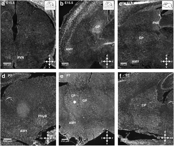
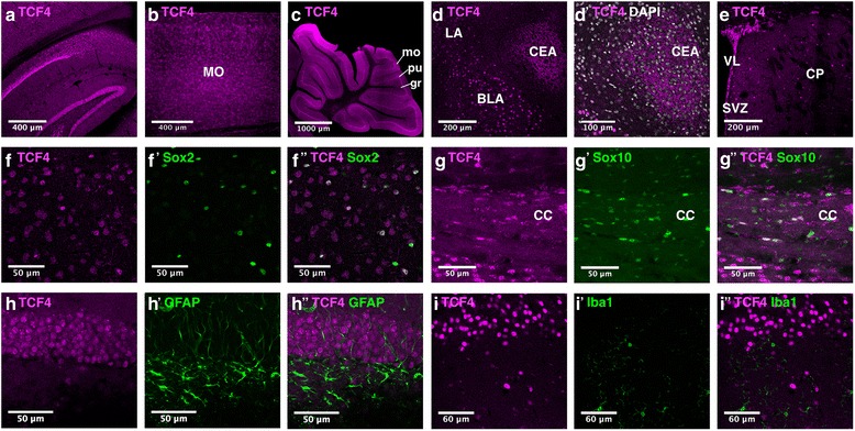
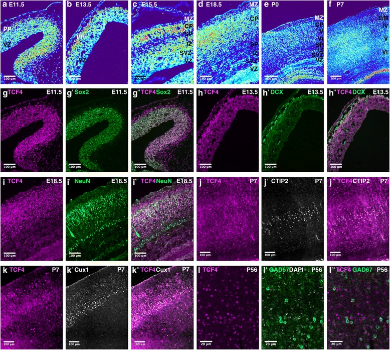
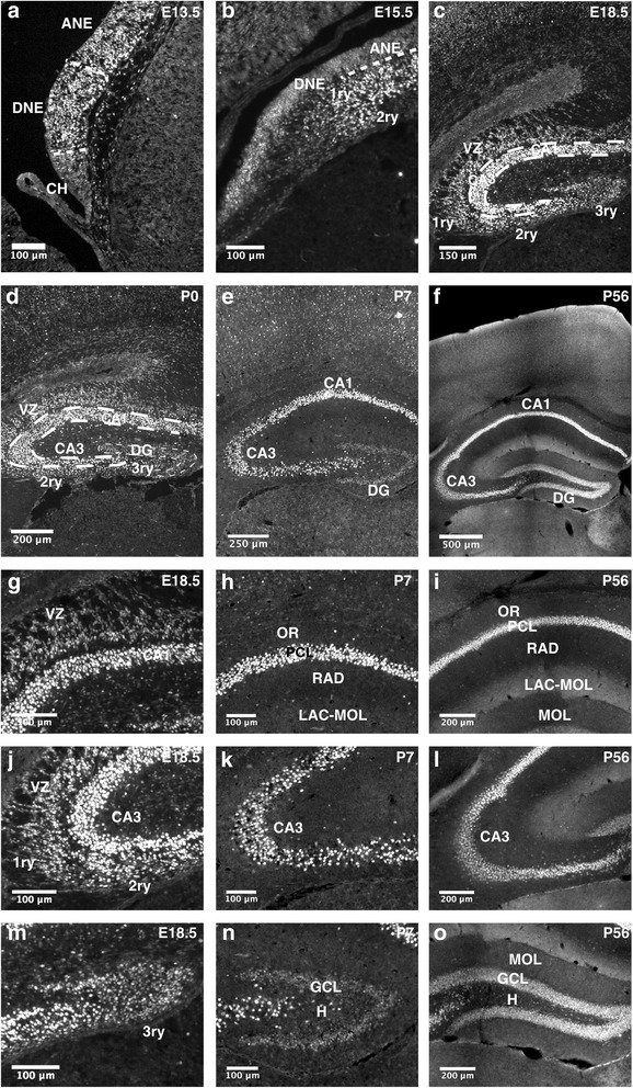
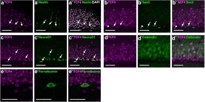
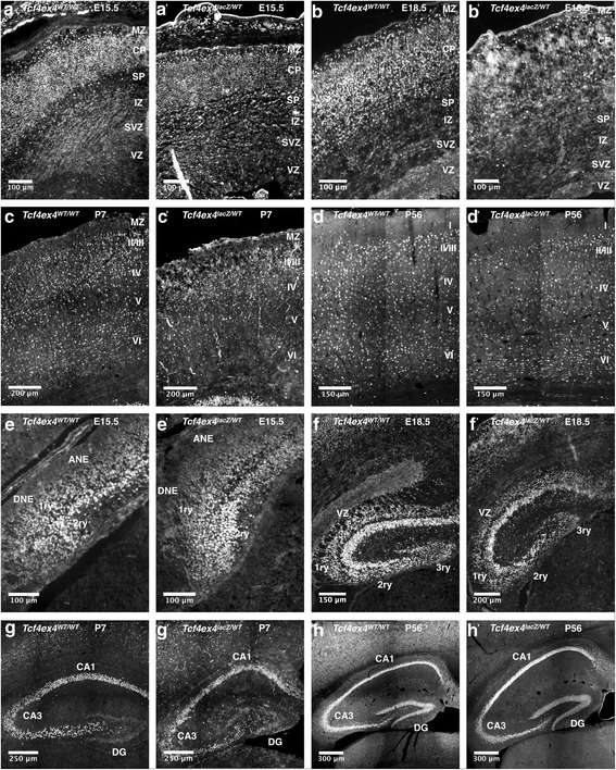
Methods: We systematically analyzed the expression pattern of TCF4 in the developing and adult mouse brain. We used immunofluorescent staining to identify candidate regions whose development and function depend on TCF4. In addition, we determined TCF4 expression in the developing rhesus monkey brain and in the developing and adult human brain through analysis of transcriptomic datasets and compared the expression pattern between species. Finally, we morphometrically and histologically analyzed selected brain structures in Tcf4-haploinsufficient mice and compared our morphometric findings to neuroanatomical findings in PTHS patients.

Results: TCF4 is broadly expressed in cortical and subcortical structures in the developing and adult mouse brain. The TCF4 expression pattern was highly similar between humans, rhesus monkeys, and mice. Moreover, Tcf4 haploinsufficiency in mice replicated structural brain anomalies observed in PTHS patients.

Conclusion: Our data suggests that TCF4 is involved in the development and function of multiple brain regions and indicates that its regulation is evolutionary conserved. Moreover, our data validate Tcf4-haploinsufficient mice as a model to study the neurodevelopmental basis of PTHS.

Keywords: Neurodevelopment; Pitt-Hopkins syndrome; Schizophrenia; TCF4.


Conflict of interest statement

The authors declare that they have no competing interests.Springer Nature remains neutral with regard to jurisdictional claims in published maps and institutional affiliations.

Figures

Fig. 1
Fig. 1
TCF4 expression at different developmental stages and validation of antibody specificity. a Western blot analysis of HEK 293T transfected with either CAG-GFP-IRES-GFP (Control), CAG-TCF4-IRES-GFP/ CAG-shTCF4-IRES-GFP (TCF4 KD), or CAG-TCF4-IRES-GFP (TCF4). Detection using the anti-TCF4 antibody revealed a clear band in the TCF4 transfected cells, which was reduced in size in the TCF4 KD (left panel). Detection using an anti-FLAG antibody resulted in bands equal in height compared to the TCF4 staining and also showed reduction upon TCF4 KD (right panel). GAPDH was used as the loading control. b, c Immunostaining using the anti-TCF4 antibody of brain tissue of TCF4 wildtype (WT) and constitutive TCF4-KO mice. dh Left-hand side of each image, TCF4 immunostaining. Right-hand side, heatmap converted image. Relative immunoreactivity scale is shown in the upper right corner: red and blue represent high and low immunoreactivity, respectively. Abbreviations: AMY, amygdala; cn, cortical neuroepithelium; CP, caudoputamen; Cx, cortex; CxP, cortical plate; DG, dentate gyrus; H, hippocampal formation; hn, hippocampal neuroepithelium; LGE, lateral ganglionic eminence; MGE, medial ganglionic eminence; pvh, paraventricular hypothalamus
Fig. 2
Fig. 2
TCF4 expression in subcortical areas during neuronal development. TCF4 is expressed in developing hypothalamic nuclei (a), the globus pallidus (b, c, e), the amygdala (b, c, d, e), and the pallidal neuroepithelium (c). TCF4 immunoreactivity is also observed in the developing caudoputamen (e, f). Abbreviations: AMY, amygdala; PHyB, basal peduncular hypothalamus; CP, caudoputamen; GP, globus pallidus; MGE, medial ganglionic eminence; PVN, paraventricular hypothalamic nucleus; PNE, pallidal neuroepithelium
Fig. 3
Fig. 3
Comparison of developmental and adult TCF4 expression. a Tabular overview of TCF4 expression during neural development. +++, high. ++, moderate. +, low. -, not detected. Hatched cell: structure not defined at developmental stage. Empty cell: expression not determined. b TCF4 expression in selected brain areas during non-human primate neural development from E40–0 months. Microarray data of six probes is grouped by brain regions and arranged by ascending age per region (blow-up) (The NIH Blueprint Non-Human Primate (NHP) Atlas, http://www.blueprintnhpatlas.org). c TCF4 expression in selected brain areas during human neural development from post-conception week 15–21. Microarray data of three probes are grouped by brain regions and aligned by ascending age per region (BrainSpan: Atlas of the Developing Human Brain [Internet]. Funded by ARRA Awards 1RC2MH089921-01, 1RC2MH090047-01, and 1RC2MH089929-01. © 2011. Available from: http://www.brainspan.org/lcm/search?search_type=user_selections). d Tabular overview of TCF4 expression in the adult mouse brain (P56). e Brain region-specific TCF4 expression in the adult human brain [23]
Fig. 4
Fig. 4
TCF4 expression in regions and non-neuronal cell types of the adult brain. In the adult brain, TCF4 is expressed in the hippocampus (A), the cortex (A, B), the cerebellum (C), the amygdaloid complex (DD’) and the caudoputamen (E). TCF4 immunoreactivity is observed in astrocytes (FF”, HH”) and oligodendrocytes (GG”) but not in microglial cells (II”). Abbreviations: BLA basolateral amydala nucleus; CC, corpus callosum; CEA, central amygdala nucleus; CP, caudoputamen; gr, granule cell layer; LA, lateral amygdala. MO, somatomotor areas; mo, molecular cell layer; pu, purkinje cell layer nucleus; SVZ, subventricular zone; VZ, ventricular zone
Fig. 5
Fig. 5
TCF4 expression during corticogenesis. (AF) Heatmap converted TCF4 immunoreactivity at different stages of cortical development. TCF4 is expressed in all layers of the neocortex with particular high intensity in the expanding cortical plate during development (GI”). TCF4 is expressed in Sox2-positive ventricular zone cells (GG”), DCX-positive immature neurons (HH”), and NeuN-positive neurons (II”). TCF4 co-stainings with layer V/VI neuron marker CTIP2 (JJ”) or layer II/III/IV neuron marker Cux1 (K-K”) at P7. TCF4 is expressed in layers II-VI but only individual TCF4-positive cells co-localize with the layer markers. TCF4 is expressed in GAD67-positive interneurons (LL”). Abbreviations: CP, cortical plate; IZ, intermediate zone; MZ, marginal zone; PP, preplate; SP, subplate; SVZ, subventricular zone; VZ, ventricular zone
Fig. 6
Fig. 6
TCF4 expression during hippocampal development. af Overview images of the hippocampal formation at different developmental stages. TCF4 is expressed throughout pre- and postnatal development of the hippocampus. gi TCF4 expression in the CA1 region of the hippocampus. At E18.5, TCF4 is expressed in radially migrating pyramidal progenitor cells and in the forming pyramidal cell layer. Postnatally, TCF4 is expressed in the pyramidal cell layer and in scattered cells in the stratum oriens and radiatum. jl At E18.5, TCF4 is expressed in dentate progenitor cells that migrate to the subpial zone (primary and secondary matrix) and in the forming pyramidal cell layer of the CA3 region. At postnatal stages P7 and P56, TCF4 is expressed in the pyramidal cell layer and in scattered cells of the strata oriens and radiatum. mo At E18.5, TCF4 is expressed in dentate progenitor cells that accumulate in the hippocampal fissure (tertiary matrix). Postnatally, TCF4 is expressed in the granular cell layer of the dentate gyrus and in individual cells of the hilus. Abbreviations: 1ry, primary matrix; 2ry, secondary matrix; 3ry, tertiary matrix; ANE, ammonic neuroepithelium; CH, cortical hem; DG, dentate gyrus; DNE, dentate neuroepithelium; GCL, granular cell layer; H, hilus; LAC-MOL, stratum lacunosum-moleculare; MOL, molecular layer; OR, stratum oriens; PCL, pyramidal cell layer; RAD, stratum radiatum; SVZ, subventricular zone
Fig. 7
Fig. 7
TCF4 expression in different cell types in the adult dentate gyrus. AA” TCF4 is expressed in Nestin-positive radial glia-like cells. BB” TCF4 is partially expressed in Sox2-positive cells. CC” TCF4 is expressed in NeuroD-positive immature neurons, DD” TCF4 is expressed in Calbindin-positive mature neurons, and EE” in Parvalbumin-positive interneurons. Arrowheads mark TCF4 negative/cell type marker positive cells. Scale bars are 40 μm
Fig. 8
Fig. 8
PTHS-associated brain anomalies in TCF4-haploinsufficient mice. Hypoplastic corpus callosum in Tcf4-haploinsufficient PTHS patients at the age of 3 years (B) and 7 years (D). A, C Age-matched references. E Small hippocampus in PTHS patient at the age of 7 years. Tcf4-haploinsufficient mice (F’, G’) show agenesis of the corpus callosum and reduced hippocampal volume (H) compared to WT mice (F, G). Cortical diameter was determined along the rostro-caudal axis (I). Representative images of the cortical thickness of Tcf4ex4WT/WT mice (J, L) and Tcf4ex4lacZ/WT mice (K, M). Cortical diameter of Tcf4ex4lacZ/WT mice was significantly decreased compared to Tcf4ex4WT/WT mice in all analyzed cortical regions. The results are displayed as mean in microns ± SD. NP Scale bars 500 μm. The DG area was measured in six representative slices spanning a distance of 240 μm. Abbreviations: PTLp, posterior parietal association area; SSp-bfd, primary somatosensory area-barrel field; SSp-tr, primary somatosensory area-trunk; Rost, rostral; Caud, caudal
Fig. 9
Fig. 9
Comparison of TCF4 protein expression at different stages of cortical and hippocampal development between Tcf4ex4WT/WT and Tcf4ex4lacZ/WT mice. AD’ Immunostaining using the anti-TCF4 antibody on brain tissue of Tcf4ex4WT/WT and Tcf4exlacZ/WT animals during cortical development and in the adult. EH’ Immunostaining using the anti-TCF4 antibody on brain tissue of Tcf4ex4WT/WT and Tcf4ex4lacZ/WT animals during hippocampus formation and in the adult. Abbreviations: ANE, ammonic neuroepithelium; CP, cortical plate; DG, dentate gyrus; DNE, dentate neuroepithelium; IZ, intermediate zone; MZ, marginal zone; PP, preplate; SP, subplate; SVZ, subventricular zone; VZ, ventricular zone; 1ry, primary matrix; 2ry, secondary matrix; 3ry, tertiary matrix

References

    1. Massari ME, Murre C. Helix-Loop-Helix proteins: regulators of transcription in eucaryotic organisms. Mol Cell Biol. 2000;20:429–440. doi: 10.1128/MCB.20.2.429-440.2000. - DOI - PMC - PubMed
    1. Amiel J, Rio M, de Pontual L, Redon R, Malan V, Boddaert N, Plouin P, Carter NP, Lyonnet S, Munnich A, Colleaux L. Mutations in TCF4, encoding a class I basic helix-loop-helix transcription factor, are responsible for Pitt-Hopkins syndrome, a severe epileptic encephalopathy associated with autonomic dysfunction. Am J Hum Genet. 2007;80:988–993. doi: 10.1086/515582. - DOI - PMC - PubMed
    1. Zweier C, Peippo MM, Hoyer J, Sousa S, Bottani A, Clayton-Smith J, Reardon W, Saraiva J, Cabral A, Gohring I, et al. Haploinsufficiency of TCF4 causes syndromal mental retardation with intermittent hyperventilation (Pitt-Hopkins syndrome) Am J Hum Genet. 2007;80:994–1001. doi: 10.1086/515583. - DOI - PMC - PubMed
    1. Schizophrenia Psychiatric Genome-Wide Association Study C Genome-wide association study identifies five new schizophrenia loci. Nat Genet. 2011;43:969–976. doi: 10.1038/ng.940. - DOI - PMC - PubMed
    1. Stefansson H, Ophoff RA, Steinberg S, Andreassen OA, Cichon S, Rujescu D, Werge T, Pietilainen OP, Mors O, Mortensen PB, et al. Common variants conferring risk of schizophrenia. Nature. 2009;460:744–747. - PMC - PubMed

Publication types

MeSH terms

Supplementary concepts

LinkOut - more resources