

Toward a real-time system for temporal enhanced ultrasound-guided prostate biopsy
- PMID: 29589258
- PMCID: PMC6436916
- DOI: 10.1007/s11548-018-1749-z
Toward a real-time system for temporal enhanced ultrasound-guided prostate biopsy
Abstract
Purpose: We have previously proposed temporal enhanced ultrasound (TeUS) as a new paradigm for tissue characterization. TeUS is based on analyzing a sequence of ultrasound data with deep learning and has been demonstrated to be successful for detection of cancer in ultrasound-guided prostate biopsy. Our aim is to enable the dissemination of this technology to the community for large-scale clinical validation.
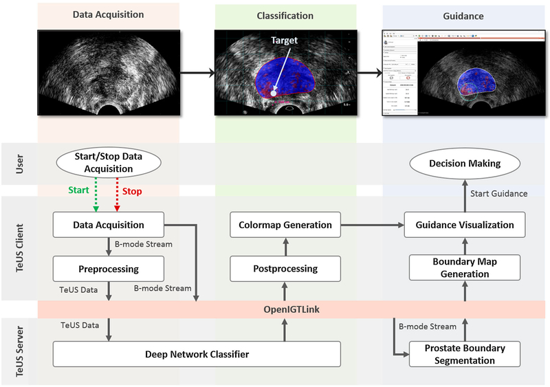
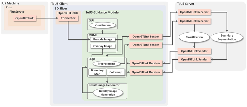
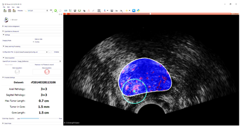
Methods: In this paper, we present a unified software framework demonstrating near-real-time analysis of ultrasound data stream using a deep learning solution. The system integrates ultrasound imaging hardware, visualization and a deep learning back-end to build an accessible, flexible and robust platform. A client-server approach is used in order to run computationally expensive algorithms in parallel. We demonstrate the efficacy of the framework using two applications as case studies. First, we show that prostate cancer detection using near-real-time analysis of RF and B-mode TeUS data and deep learning is feasible. Second, we present real-time segmentation of ultrasound prostate data using an integrated deep learning solution.
Results: The system is evaluated for cancer detection accuracy on ultrasound data obtained from a large clinical study with 255 biopsy cores from 157 subjects. It is further assessed with an independent dataset with 21 biopsy targets from six subjects. In the first study, we achieve area under the curve, sensitivity, specificity and accuracy of 0.94, 0.77, 0.94 and 0.92, respectively, for the detection of prostate cancer. In the second study, we achieve an AUC of 0.85.
Conclusion: Our results suggest that TeUS-guided biopsy can be potentially effective for the detection of prostate cancer.
Keywords: 3D slicer; Prostate cancer; Real-time biopsy guidance; Temporal enhanced ultrasound.
Conflict of interest statement
Figures
References
-
- Abadi M, Agarwal A, Barham P, Brevdo E, Chen Z, Citro C, Corrado GS, Davis A, Dean J, Devin M (2016) Tensorflow: large-scale machine learning on heterogeneous distributed systems. arXiv preprint arXiv:1603.04467
-
- Ahmed HU, Bosaily AES, Brown LC, Gabe R, Kaplan R, Parmar MK, Collaco-Moraes Y, Ward K, Hindley RG, Freeman A (2017) Diagnostic accuracy of multi-parametric MRI and TRUS biopsy in prostate cancer (PROMIS): a paired validating confirmatory study. Lancet 389(10071):815–822 - PubMed
-
- Anas EMA, Nouranian S, Mahdavi SS, Spadinger I, Morris WJ, Salcudean SE, Mousavi P, Abolmaesumi P (2017) Clinical target-volume delineation in prostate brachytherapy using residual neural networks In: International conference on medical image computing and computer-assisted intervention. Springer, pp 365–373
-
- Azizi S, Bayat S, Yan P, Tahmasebi A, Nir G, Kwak JT, Xu S, Wilson S, Iczkowski KA, Lucia MS, goldenberg L, Salcudean SE, Pinto P, Wood B, Abolmaesumi P, Mousavi P (2017) Detection and grading of prostate cancer using temporal enhanced ultrasound: combining deep neural networks and tissue mimicking simulations. Int J Comput Assist Radiol Surg 12(8):1293–1305 - PMC - PubMed
MeSH terms
Grants and funding
LinkOut - more resources
Full Text Sources
Other Literature Sources
Medical
