. 2018 Mar 28;13(3):e0193583.
doi: 10.1371/journal.pone.0193583. eCollection 2018.

Humanin (HN) and glucose transporter 8 (GLUT8) in pregnancies complicated by intrauterine growth restriction

Affiliations

Humanin (HN) and glucose transporter 8 (GLUT8) in pregnancies complicated by intrauterine growth restriction

Carla Janzen et al. PLoS One. .

Abstract

Background: Intrauterine growth restriction (IUGR) results from a lack of nutrients transferred to the developing fetus, particularly oxygen and glucose. Increased expression of the cytoprotective mitochondrial peptide, humanin (HN), and the glucose transporter 8, GLUT8, has been reported under conditions of hypoxic stress. However, the presence and cellular localization of HN and GLUT8 in IUGR-related placental pathology remain unexplored. Thus, we undertook this study to investigate placental expression of HN and GLUT8 in IUGR-affected versus normal pregnancies.

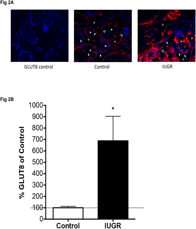
Results: We found 1) increased HN expression in human IUGR-affected pregnancies on the maternal aspect of the placenta (extravillous trophoblastic (EVT) cytoplasm) compared to control (i.e. appropriate for gestational age) pregnancies, and a concomitant increase in GLUT8 expression in the same compartment, 2) HN and GLUT8 showed a protein-protein interaction by co-immunoprecipitation, 3) elevated HN and GLUT8 levels in vitro under simulated hypoxia in human EVT cells, HTR8/SVneo, and 4) increased HN expression but attenuated GLUT8 expression in vitro under serum deprivation in HTR8/SVneo cells.

Conclusions: There was elevated HN expression with cytoplasmic localization to EVTs on the maternal aspect of the human placenta affected by IUGR, also associated with increased GLUT8 expression. We found that while hypoxia increased both HN and GLUT8, serum deprivation increased HN expression alone. Also, a protein-protein interaction between HN and GLUT8 suggests that their interaction may fulfill a biologic role that requires interdependency. Future investigations delineating molecular interactions between these proteins are required to fully uncover their role in IUGR-affected pregnancies.


Conflict of interest statement

Competing Interests: The authors have declared that no competing interests exist.

Figures

Fig 1
Fig 1. mRNA and protein levels of GLUT8 are increased in IUGR-affected pregnancies.
(A) Real-time quantitative RT-PCR analysis of placental GLUT8 expression. IUGR-affected placentas (n = 22) were assayed against gestational age-matched, control placentas (n = 22) for the maternal and fetal sides. Relative quantification of PCR products was based on the CT value differences between target and the housekeeping gene using the comparative CT method (Eukaryotic 18S rRNA) was used as an internal control. (*** = p<0.001). (B) Western blot analysis of GLUT8 in placental biopsies obtained from women with normal pregnancy (n = 22) or IUGR-affected pregnancy, by abnormal prenatal ultrasound (n = 22). Vinculin was used as the loading control. Representative blots are shown. (** = p<0.01). The arrow pointing at the dark band at approximately 130 kDa indicates Vinculin. The arrow pointing at the dark band at approximately 51 kDa indicates GLUT8. Sample label abbreviations are as follows: "CM" is control maternal side, "CF" is control fetal side, "IM" is IUGR maternal side, and "IF" is IUGR fetal side. The number following the sample abbreviation indicates sample ID. The same number on multiple labels indicates the same sample. Brain was used as a positive control for GLUT8.
Fig 2
Fig 2. Increased immunofluorescence of GLUT8 in IUGR-affected placentas.
(A) Immunoflourescence staining of GLUT8 (red) and DAPI (blue) of placental tissue. Left: negative control performed with non-isotype IgG to measure the level of non-specific background signal. Middle: GLUT8 staining in the normal placenta. Right: GLUT8 staining of the IUGR-affected placenta. Plus sign (+) indicates intervillous space. Asterisk (*) shows villous core. Line arrows show syncytiotrophoblast. Hollow arrows show cytotrophoblast. All images taken at 40X magnification. (B) Relative quantitation of GLUT8 immunofluorescence staining of GLUT8 in the normal and IUGR-affected human placenta. (* = p<0.05). Images were captured with Nikon E-600 microscope (Nikon, NY), equipped with a cooled charge-coupled device (CCD) camera (Cool SNAP HQ Monochrome, Roper Scientific, AZ). There were 5 control and 5 IUGR placentas, full thickness section for each placenta. 20 fields were randomly selected for each section. Images were acquired at 12-bit gray level resolution and displayed in psuedocolor (monochrome) for analysis with GLUT8 represented by Alexa Fluor 488-conjugated donkey anti-rabbit IgG. The Metamorph image analysis system software (Molecular Devices, CA) was used to measure integrated optical density for GLUT8 expression.
Fig 3
Fig 3. Increased immunohistochemical staining of HN in IUGR-affected placentas.
(A) Immunohistochemical staining of humanin (HN) in human placental tissue at gestational age of 38 weeks. Humanin was stained in brown. The left column shows staining of the stem villi EVT of placental tissue, and the right column shows staining of the maternal surface EVT of placental tissue. The top row of both columns is negative control performed with non-isotype IgG. The middle row of both columns is placenta from normal pregnancies. The bottom row of both columns is IUGR. 40x magnification for all images. (B) Immunohistochemical staining in normal (control) human placental tissue showing four different markers of same placental sample (serial sections) at 38 weeks gestational age. The top row is the non-isotype IgG control for each of the markers labeled. The bottom row from left to right is staining of AE1/AE3 (trophoblast marker), GLUT8, Humanin, and lysozyme (lysosomal marker). The presence of EVT on the right portion of each image is supported by diffuse cytoplasmic staining of AE1/AE3, while the endometrial stromal cells within the decidua on the left portion of each image are negative for AE1/AE3. Each of the four images on the bottom row shows the decidua on the left side and maternal surface EVT on the right side of the image. Arrows point to maternal surface EVT and triangles indicate decidual stromal cells. All images were taken at 20x magnification.
Fig 4
Fig 4. Immunoprecipitation (IP) assays reveal association between endogenous humanin with Glut8 in HTR-8 cells.
(A) HTR8/SVneo protein was immunoprecipitated with Glut8 antibody then the bound protein was analyzed by western blot with humanin antibody. (B) HTR8/SVneo protein lysate was immunoprecipitated with humanin antibody then analyzed by western blot with glut8 antibody. In both the cases Rabbit non-isotype IgG was used as negative control (-C) and in positive control (+C), both the immunoprecipitation and western blot analyses were done using the same antibody (either humanin or Glut8 antibody).
Fig 5
Fig 5. Immunocytochemical staining showing localization of HN and GLUT8 in HTR8/SVneo cells.
The top rows show localization of GLUT8. On the top row, the left image is the non-isotype IgG control, the left middle is GLUT8 (green) staining taken using a filter for GLUT8 alone, the right middle is DAPI (blue) taken using a filter for DAPI alone, and the right image is GLUT8 (green) and DAPI (blue) combined. The 3 rightmost images are of the same area. The bottom rows show images of humanin. On the bottom row, the left image is the non-isotype IgG control, the left middle is Humanin (green) staining using a filter for Humanin alone, the right middle is DAPI (blue) using a filter for DAPI alone, and the right image is Humanin (green) and DAPI (blue) combined. The three rightmost images are of the same area. All images have 40x magnification. Scale bar = 50 μm.
Fig 6
Fig 6. CoCl2-stimulated hypoxia induces HN and GLUT8 expression in HTR8/SVneo cells, and serum-deprived HTR8/SVneo cells up-regulates HN levels but down-regulates GLUT8 levels.
(A) HN and GLUT8 levels were determined by Western blot analysis at 4, 8, and 24 hours of incubation of HTR8/SVneo cells in 250 and 500 μM CoCl2–supplemented complete media. Representative blots are shown. β-actin and vinculin were used as loading controls for HN and GLUT8, respectively (n = 3, * = p<0.05, ** = p<0.01, and *** = p<0.001). (B) HN and GLUT8 expression was also monitored in HTR8/SVneo cells at 8, 24 and 48 hours of cultivation in serum-supplemented (FBS (+)) and serum-free media (FBS (-)). Representative blots are shown. β-actin and vinculin were used as the loading controls for HN and GLUT8, respectively. (n = 3, * = p<0.05 and ** = p<0.01).
Fig 7
Fig 7. Primary human trophoblasts exposed to serum starvation.
Top row is GLUT8 staining (green) of human primary villous trophoblast cells. Bottom row is humanin staining (green) of human primary villous trophoblast cells. All images show DAPI staining of nuclei (blue). From left to right: non-isotype IgG control, cells grown for 8 hours with serum, cells grown for 8 hours without serum, cells grown for 24 hours with serum, cells grown for 24 hours without serum. All images taken at 40x magnification. Scale bar = 50 μm.

References

    1. Saleem T, Sajjad N, Fatima S, Habib N, Ali SR, Qadir M. Intrauterine growth retardation—small events, big consequences. Italian journal of pediatrics. 2011;37:41 Epub 2011/09/09. doi: 10.1186/1824-7288-37-41 . - DOI - PMC - PubMed
    1. Biri A, Bozkurt N, Turp A, Kavutcu M, Himmetoglu O, Durak I. Role of oxidative stress in intrauterine growth restriction. Gynecologic and obstetric investigation. 2007;64(4):187–92. Epub 2007/08/01. doi: 10.1159/000106488 . - DOI - PubMed
    1. Caniggia I, Winter J, Lye SJ, Post M. Oxygen and placental development during the first trimester: implications for the pathophysiology of pre-eclampsia. Placenta. 2000;21 Suppl A:S25–30. Epub 2000/06/01. . - PubMed
    1. Genbacev O, Zhou Y, Ludlow JW, Fisher SJ. Regulation of human placental development by oxygen tension. Science. 1997;277(5332):1669–72. Epub 1997/09/12. . - PubMed
    1. Klein LE, Cui L, Gong Z, Su K, Muzumdar R. A humanin analog decreases oxidative stress and preserves mitochondrial integrity in cardiac myoblasts. Biochemical and biophysical research communications. 2013;440(2):197–203. Epub 2013/08/30. doi: 10.1016/j.bbrc.2013.08.055 . - DOI - PMC - PubMed

Publication types

MeSH terms

Substances